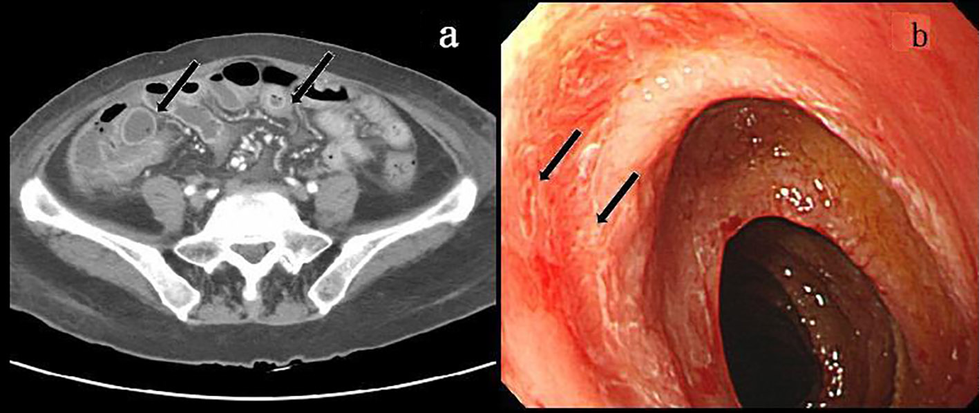
Figure 1. Pre-treatment contrast-enhanced abdominal CT showed wall-thickening of the segmental small bowel (arrow) (a), and pre-treatment enteroscopy showed hyperemic mucosal change with discrete ulcers (arrow) at the jejunum (b).
| Gastroenterology Research, ISSN 1918-2805 print, 1918-2813 online, Open Access |
| Article copyright, the authors; Journal compilation copyright, Gastroenterol Res and Elmer Press Inc |
| Journal website http://www.gastrores.org |
Case Report
Volume 10, Number 3, June 2017, pages 193-195
A Case With Vitamin D Deficiency-Induced Cytomegalovirus Enteritis Presenting as Bowel Pseudo-Obstruction
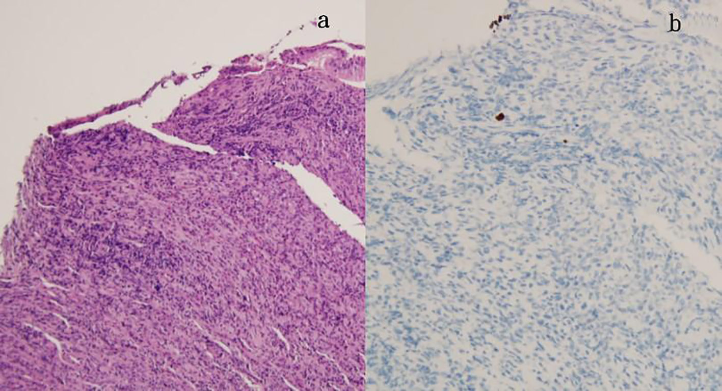
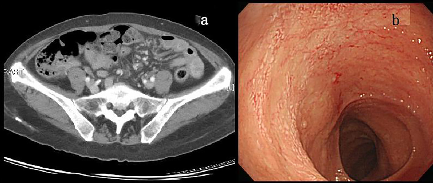
Figures