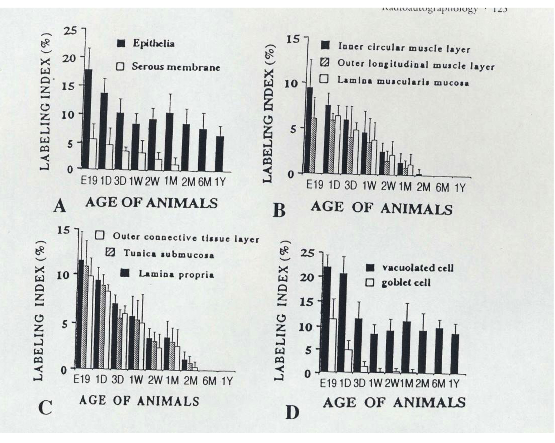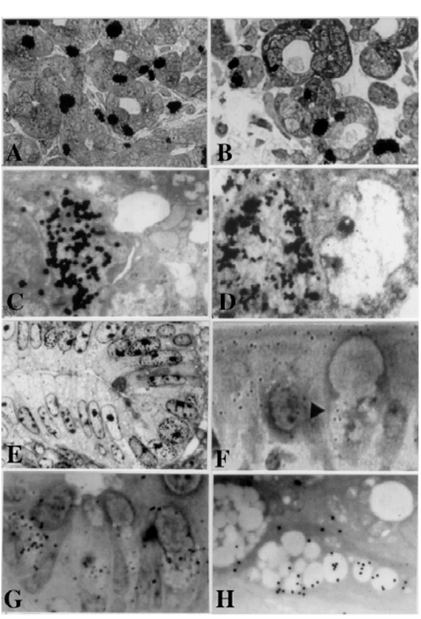
Figure 1. From Nagata, T.: Radioautographology, General and Special. In, Progr. Histochem. Cytochem. Vol. 37, No. 2, p. 118, 2002, Figure 5. LM and EM RAG of the digestive organs. Urban & Fischer, Jena, Germany. (A) LM RAG of the submandibular gland of male mouse embryonic day 19 labeled with 3H-thymidine consisted with the glandular acini and duct system (× 500). The duct system was composed of juxtaacinar cells (JA), intercalated duct cells (ICD) and striated duct cells (ICD). Many labeled developing acinar cells (AC), JA and ICD cells were observed. (B) LM RAG of the submandibular gland at postnatal day 3, labeled with 3H-thymidine (× 500). There were more JA cells and secretory granules than those of former stage (Fig. 1A). (C) EM RAG of an ICD cell of a mouse at postnatal day 3, labeled with 3H-thymidine observed by electron microscopy (× 10,000). Many silver grains are observed over the nucleus of an ICD. (D) EM RAG of the esophageal epithelial cells of a newborn mouse at postnatal day 1, labeled with 3H-thymidine (× 10,000). Many silver grains are observed over one of the nuclei at left. (E) LM RAG of the colonic epithelial cells of a mouse embryo at fetal day 19, labeled with 3H-thymidine (× 800). Many silver grains are observed over the nuclei of several epithelial cells in the bottom of the crypt. (F) LM RAG of the ileum epithelial cells labeled with 3H-glucosamine of an old mouse at postnatal 6 months (× 1,000). Many silver gains are localized over the Golgi region of the three goblet cells as well as over the cytoplasm of several absorptive columnar epithelial cells. (G) LM RAG of the colonic epithelial cells of a mouse at postnatal month 1, labeled with 35SO4 in vitro and radioautographed (× 1,000). (H) EM RAG of a goblet cell in the deeper crypt of the colonic epithelial cells of an adult mouse after injection of 35SO4 and radioautographed (× 4,800). Many silver grains are observed over the Golgi region and mucous droplets of the goblet cell, demonstrating the incorporation of radiosulfate into sulfomucins.