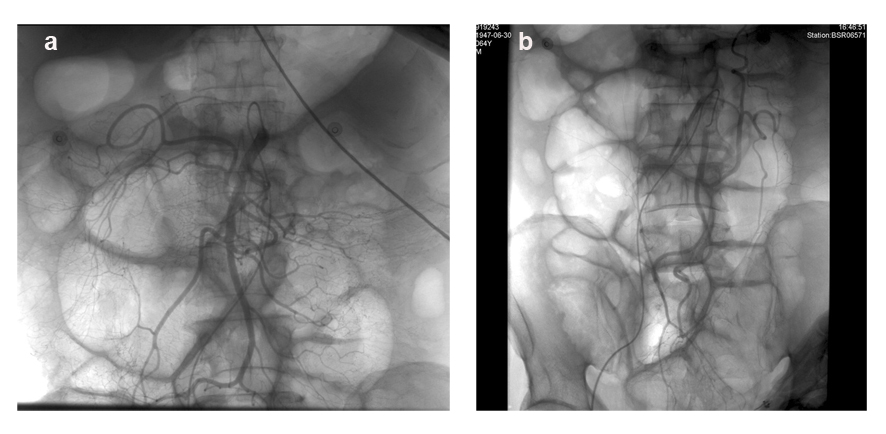
Figure 1. DSA of superior mesenteric artery and inferior mesenteric artery did not reveal any vascular lesions.
| Gastroenterology Research, ISSN 1918-2805 print, 1918-2813 online, Open Access |
| Article copyright, the authors; Journal compilation copyright, Gastroenterol Res and Elmer Press Inc |
| Journal website http://www.gastrores.org |
Case Report
Volume 7, Number 2, April 2014, pages 73-77
Hematochezia From Splenic Arterial Pseudoaneurysm Ruptured Into Pancreatic Pseudocyst Coexisting With Fistula to the Colon: A Case Report and Literature Review
Figures