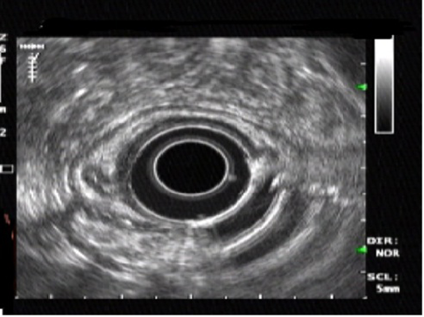
Figure 1. Endoscopic ultrasonography showing a homogeneous, hypoechoic, clearly demarcated mass in the submuscosal layer.
| Gastroenterology Research, ISSN 1918-2805 print, 1918-2813 online, Open Access |
| Article copyright, the authors; Journal compilation copyright, Gastroenterol Res and Elmer Press Inc |
| Journal website http://www.gastrores.org |
Case Report
Volume 6, Number 6, December 2013, pages 240-243
Endoscopic Removal of Granular Cell Tumors of Stomach: Case Report and Review of Literature
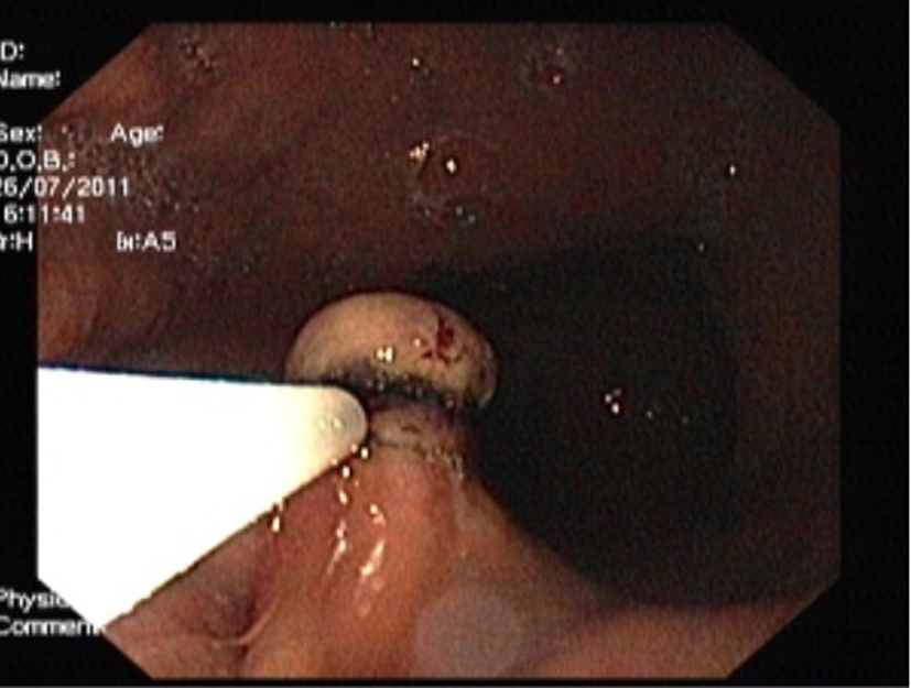
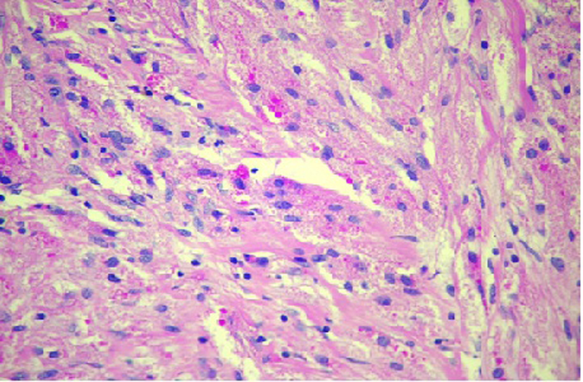
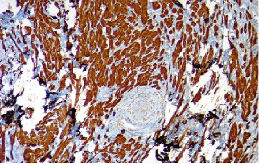
Figures