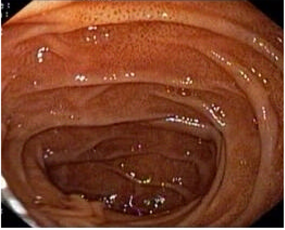
Figure 1. Esophagogastroduodenoscopy showing muliple dark pigmented spots in the second portion of the duodenum.
| Gastroenterology Research, ISSN 1918-2805 print, 1918-2813 online, Open Access |
| Article copyright, the authors; Journal compilation copyright, Gastroenterol Res and Elmer Press Inc |
| Journal website http://www.gastrores.org |
Case Report
Volume 5, Number 4, August 2012, pages 171-173
Pseudomelanosis Duodeni of Undetermined Etiology
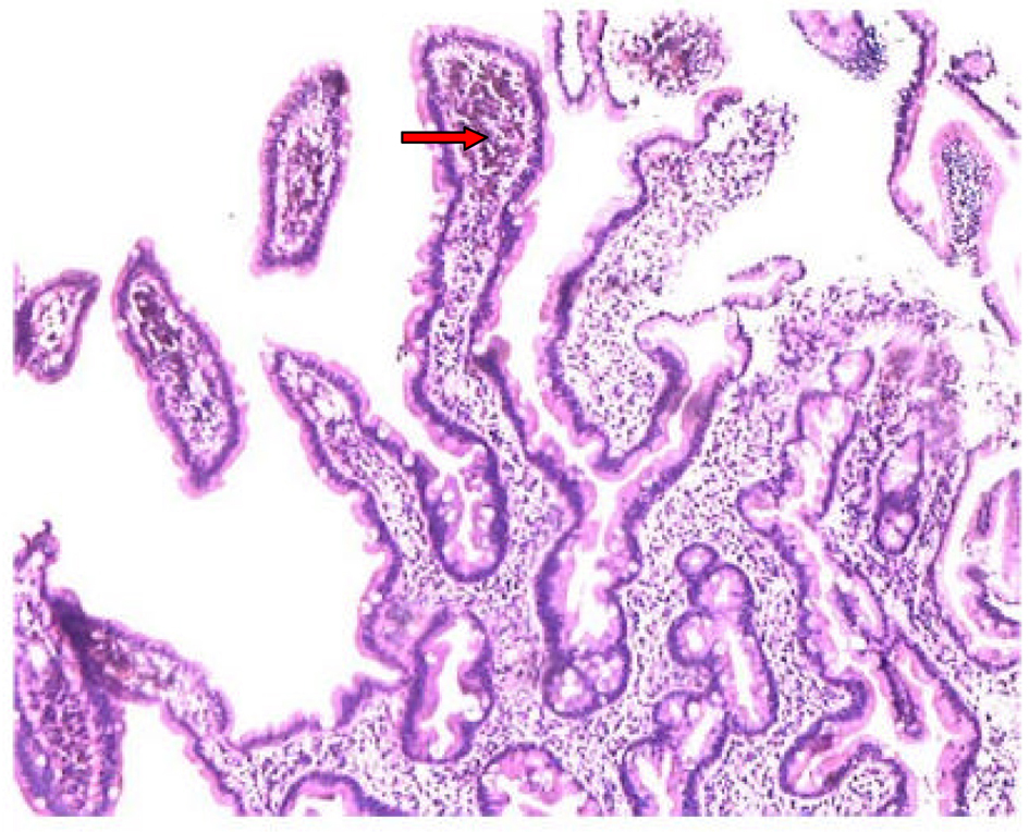
Figures