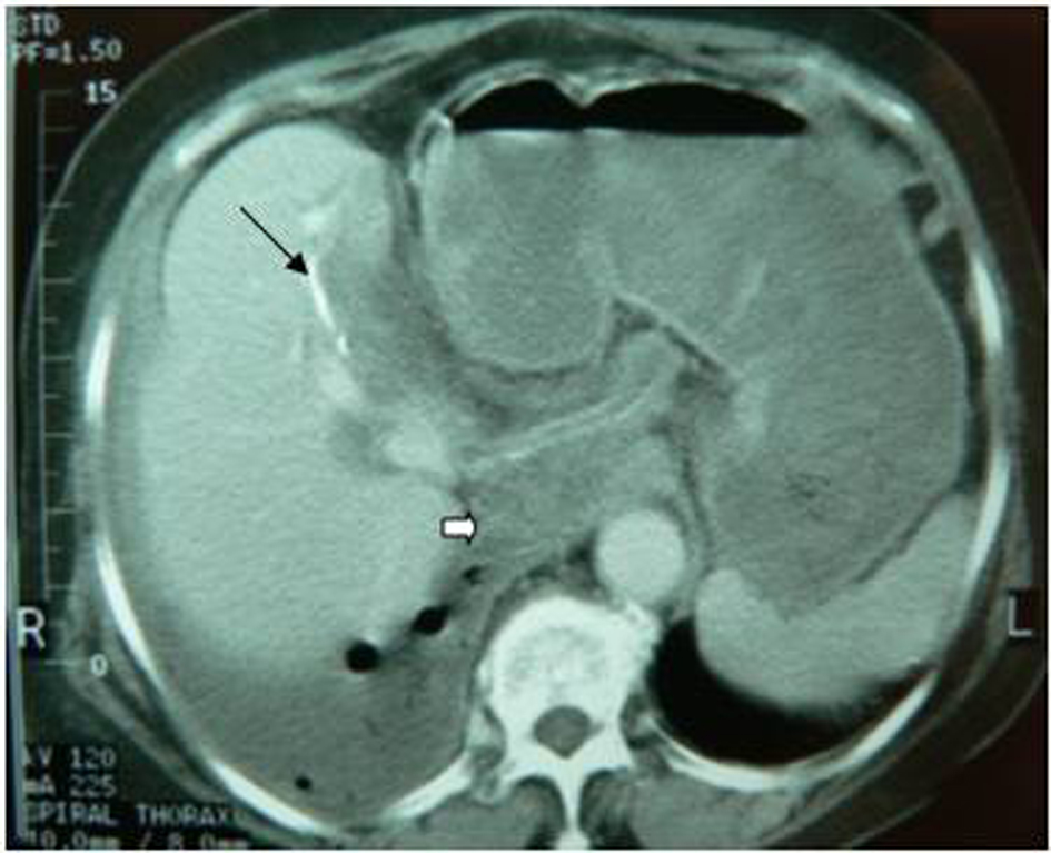
Figure 1. Transdiaphragmatic thoracic rupture. It is observed the calcified hydatid cyst wall (black arrow), daughter cysts (white arrow) and pleural effusion with presence of air.
| Gastroenterology Research, ISSN 1918-2805 print, 1918-2813 online, Open Access |
| Article copyright, the authors; Journal compilation copyright, Gastroenterol Res and Elmer Press Inc |
| Journal website http://www.gastrores.org |
Original Article
Volume 5, Number 4, August 2012, pages 139-143
Complications of Hydatid Cysts of the Liver: Spiral Computed Tomography Findings
Figures
Table
| Patients | |||||||
|---|---|---|---|---|---|---|---|
| No | 1 | 2 | 3 | 4 | 5 | 6 | 7 |
| Sex and Age | F 92 | M 70 | F 80 | F 63 | F 86 | M 65 | F 64 |
| Known history of hydatid disease | Yes | No | Yes | Yes | No | Yes | No |
| Clinical features | Acute right upper abdominal quadrant pain | Acute right upper abdominal quadrant pain; Fever | Diffuse abdominal pain Fever Abdominal wall contraction Rebound sensitivity | Diffuse abdominal pain Right thoracic pain Fever; Dyspnea | Acute right upper abdominal quadrant pain with lumbar radiation | Acute right upper abdominal quadrant pain; Fever | Acute right upper abdominal quadrant pain |
| Laboratory findings | Eosinophilia Increased amylase serum Increased direct bilirubin serum | Eosinophilia Increased direct bilirubin serum | Increased WBC count | Eosinophilia | Mild leukocytosis | leukocytosis | Increased direct bilirubin serum |
| Treatment | Surger | Surgery | Surgery | Surgery | Surgery | Surgery | Surgery |
| Outcome | Improvement | Improvement | Death | Improvement | Improvement | Improvement | Improvement |