Gastroenterology Research, ISSN 1918-2805 print, 1918-2813 online, Open Access
Article copyright, the authors; Journal compilation copyright, Gastroenterol Res and Elmer Press Inc
Journal website http://www.gastrores.org

Case Report

Volume 5, Number 2, April 2012, pages 71-73


First Case of Small Bowel Sarcomatoid Carcinoma Found by Video Capsule Endoscopy

Figures

Figure 1.
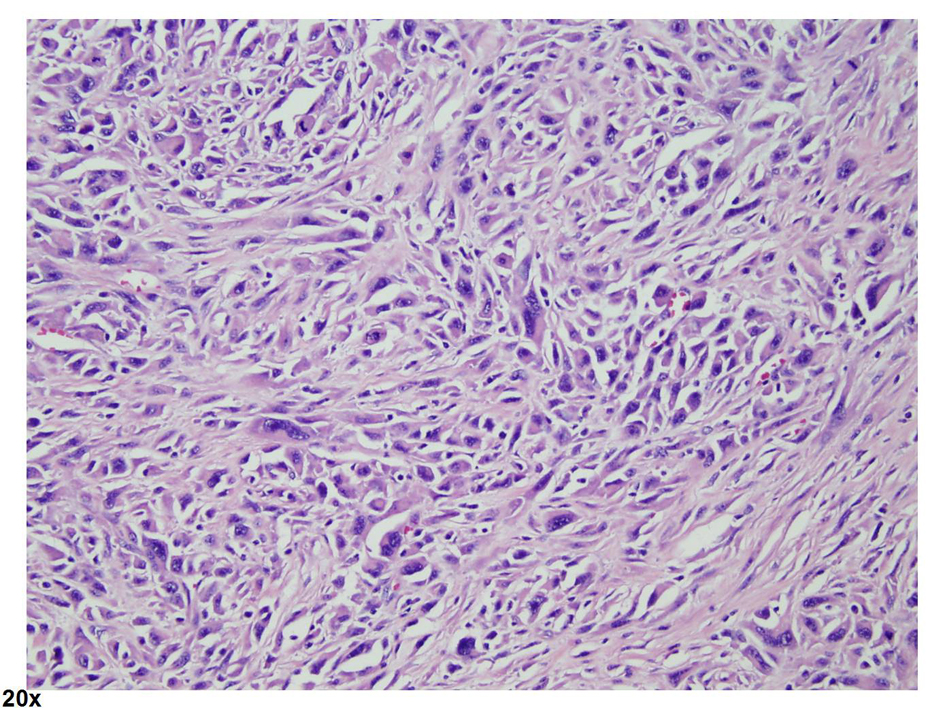
Figure 1. Hematoxylin and eosin (H and E) stain. Pleomorphic spindle cells with moderate amount of eosinophilic cytoplasm, irregular nuclear membranes, vesicular chromatin and prominent nucleoli. Numerous mitotic figures are noted. Objective magnification x 20
Figure 2.
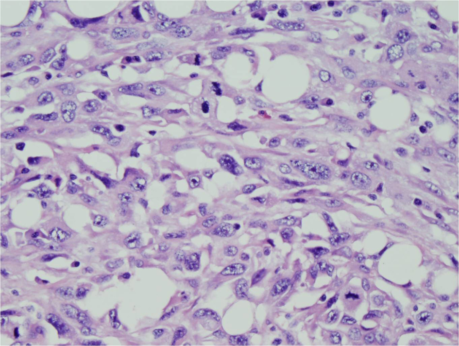
Figure 2. Hematoxylin and eosin (H and E) stain. Pleomorphic spindled to epitheliod cells with moderate amount of eosinophilic cytoplasm, vesicular chromatin and prominent nucleoli.  Numerous mitotic figures are noted. Scattered plasma cells are seen in the background.  Objective magnification x 40.
Figure 3.
Figure 3. Positive staining for CAM 5.2 immunohistochemical stain. Objective magnification x 50.
Figure 4.
Figure 4. Positive staining for Vimentin immunohistochemical stain. Objective magnification x 50.