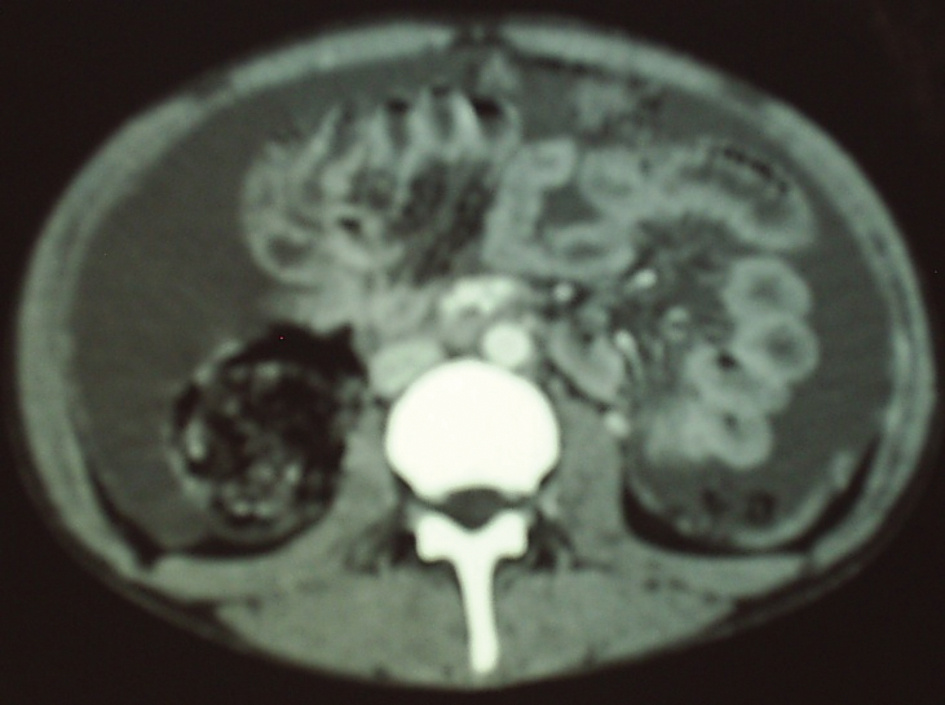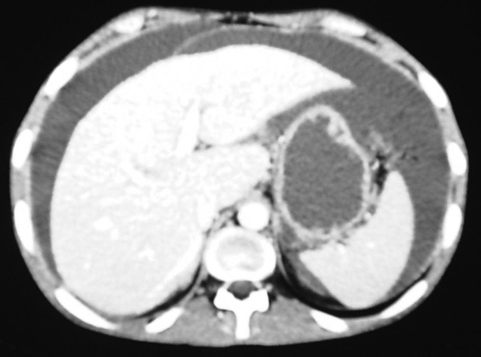
Figure 1. Ascites apperance on CT of the abdomen.
| Gastroenterology Research, ISSN 1918-2805 print, 1918-2813 online, Open Access |
| Article copyright, the authors; Journal compilation copyright, Gastroenterol Res and Elmer Press Inc |
| Journal website http://www.gastrores.org |
Case Report
Volume 5, Number 3, June 2012, pages 127-129
Endodermal Sinus Tumor Presented With Ascites: A Case Report
Figures