Gastroenterology Research, ISSN 1918-2805 print, 1918-2813 online, Open Access
Article copyright, the authors; Journal compilation copyright, Gastroenterol Res and Elmer Press Inc
Journal website http://www.gastrores.org

Original Article

Volume 4, Number 2, April 2011, pages 70-75


Alterations of Mast Cells in the Esophageal Mucosa of the Patients With Non-Erosive Reflux Disease

Figures

Figure 1.
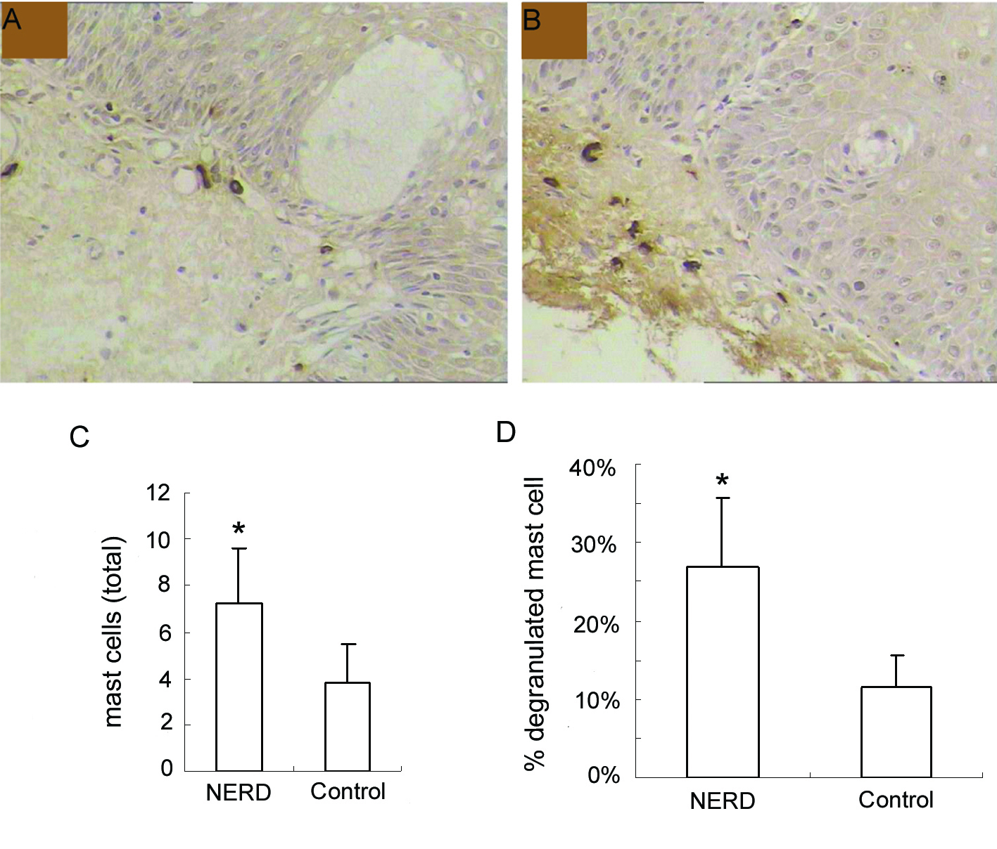
Figure 1. Immunohistochemical analysis of mast cells at the esophageal mucosa related to ‘NERD’ and ‘control’. (A), control, and (B), NERD patients, illustrate the immunohistochemical staining of mast cells (tryptase immunostain, brown cells). Slides shown (tryptase immunostain counterstained with hematoxylin × 400 magnification) are representative of common findings. The data are presented as columns displaying the means of the total number per high-power field (C) and the relative proportion of degranulated mast cells of all intramucosal mast cells (D) (HPF; magnification × 400) ± standard error (SE). *P < 0.01.
Figure 2.
Figure 2. Electron micrograph of a mast cell in the lamina propria of esophageal mucosa in NERD. The cytoplasm of the mast cell showes degranulation with multiple ghost vacuoles (arrows) and targetoid-type granules (open arrows). Original magnification × 6000. Scale bar = 2 µm.