Figure 1. Liver lesion: CT image showing large hypodense lesion in the left liver lobe (arrow).
| Gastroenterology Research, ISSN 1918-2805 print, 1918-2813 online, Open Access |
| Article copyright, the authors; Journal compilation copyright, Gastroenterol Res and Elmer Press Inc |
| Journal website http://www.gastrores.org |
Case Report
Volume 3, Number 6, December 2010, pages 293-295
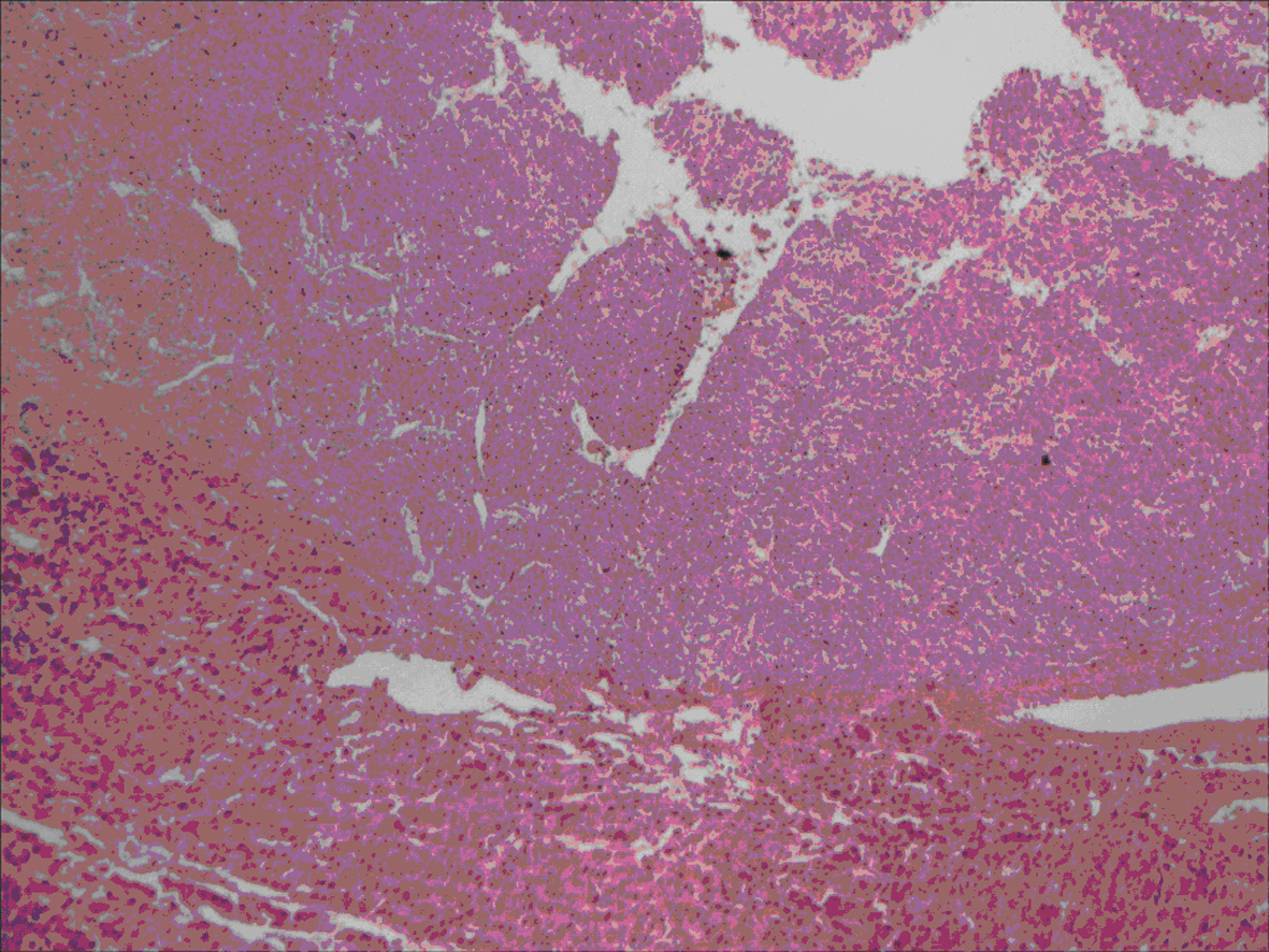
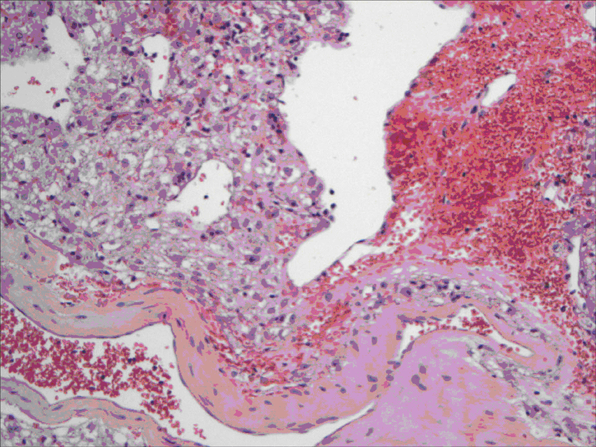
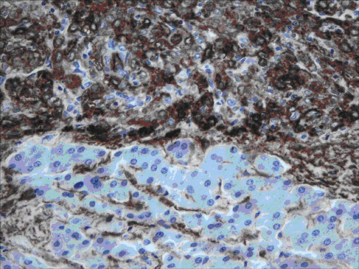
Hepatic Epithelioid Angiomyolipoma: Case Series
Figures