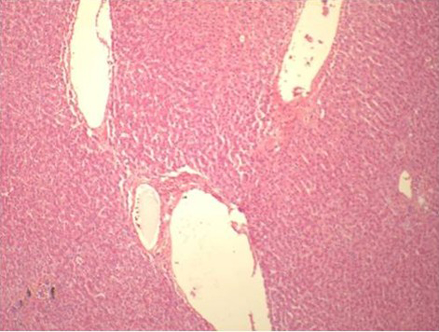
Figure 1. Liver Biopsy showing normal architecture
| Gastroenterology Research, ISSN 1918-2805 print, 1918-2813 online, Open Access |
| Article copyright, the authors; Journal compilation copyright, Gastroenterol Res and Elmer Press Inc |
| Journal website http://www.gastrores.org |
Case Report
Volume 3, Number 6, December 2010, pages 281-286
A Case of Isolated Duodenal Varices Secondary to Chronic Pancreatitis with Review of Literature
Figures
Tables
| Cirrhosis of Liver [1, 4-6] |
| Thrombotic disorders and Coagulopathies [4] |
| Schistosomiasis [7] |
| Pancreatitis and Cholangitis [3, 8] |
| Omphalophlebitis (surgical catheterization of Umbilicus in adults) [8-10] |
| Pancreatic and periportal tumors [8] |
| Mesenteric Metastasis [4, 11] |
| Surgical Procedure or Trauma [4, 11] |
| Visceral Hemangiomata (Klippel-Trenaunay-Weber syndrome) [12-16] |
| Vascular Anomalies (Occlusion of SMV by abnormal Ileocecal Artery) [12-16] |
| Retroperitoneal Fibrosis [8] |
| Part of Duodenum | Duodenal Bulb | 48 |
|---|---|---|
| 2nd portion | 12 | |
| 3rd portion | 3 | |
| 4th portion | 1 | |
| DuodenoJejunal Junction | 3 | |
| Not Specified | 38 | |
| Associated Varices | Esophagus | 25 |
| Esophagus and Stomach | 2 | |
| Esophagus, Stomach and Jejunum | 1 | |
| Not recognized | 5 | |
| Not Specified | 72 |