Gastroenterology Research, ISSN 1918-2805 print, 1918-2813 online, Open Access
Article copyright, the authors; Journal compilation copyright, Gastroenterol Res and Elmer Press Inc
Journal website http://www.gastrores.org

Case Report

Volume 3, Number 5, October 2010, pages 219-222


Thyroid Metastasis of Gastric Cancer: A Rare Occasion With Poor Prognosis

Figures

Figure 1.
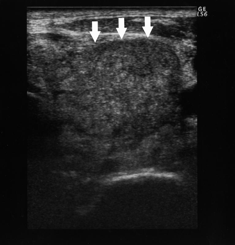
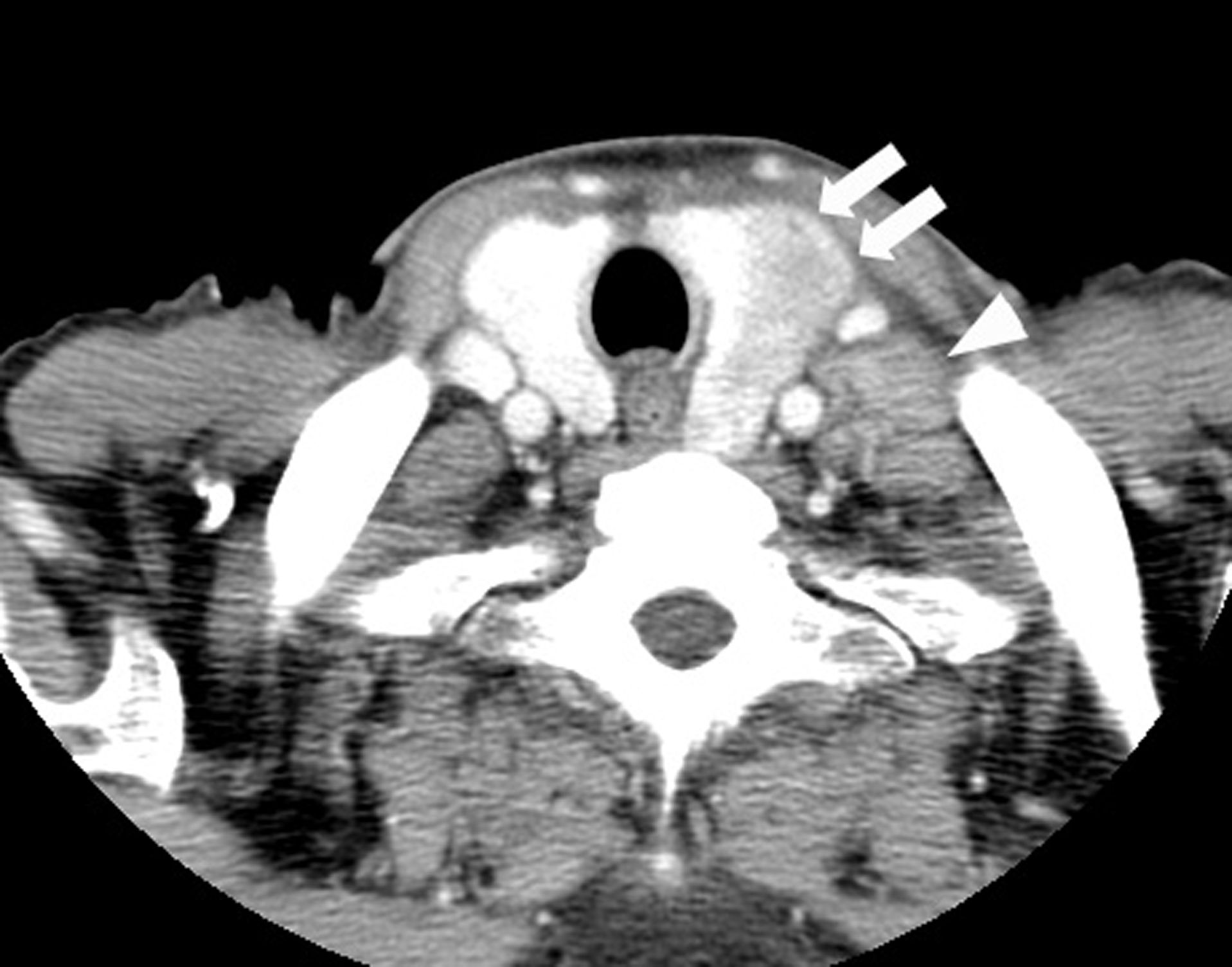
Figure 1. CT findings of the thyroid tumor. Thyroid tumor in the left lobe (arrows) and the left cervical lymph node swelling (arrowhead) are detected in contrast-enhanced CT. This thyroid tumor is a heterogeneously low-density tumor with an unclear border. The trachea is deviated to the right by this tumor.
Figure 2.
Figure 2. Ultrasonographic findings of the thyroid tumor. Ultrasonography reveals a 3.1 cm-sized tumor in the left lobe of the thyroid gland (arrows). It shows mosaic echogenecity and no calcification inside with a partially unclear border but no apparent spicular formation.
Figure 3.
Figure 3. Cytology of the specimen that was obtained by FNAB. A signet-ring cell carcinoma cell and poorly differentiated adenocarcinoma cells with high degree of cellular atypia and a high nucleus/cytoplasm ratio are found by cytology (Papanicolau stain; x 200).
Figure 4.
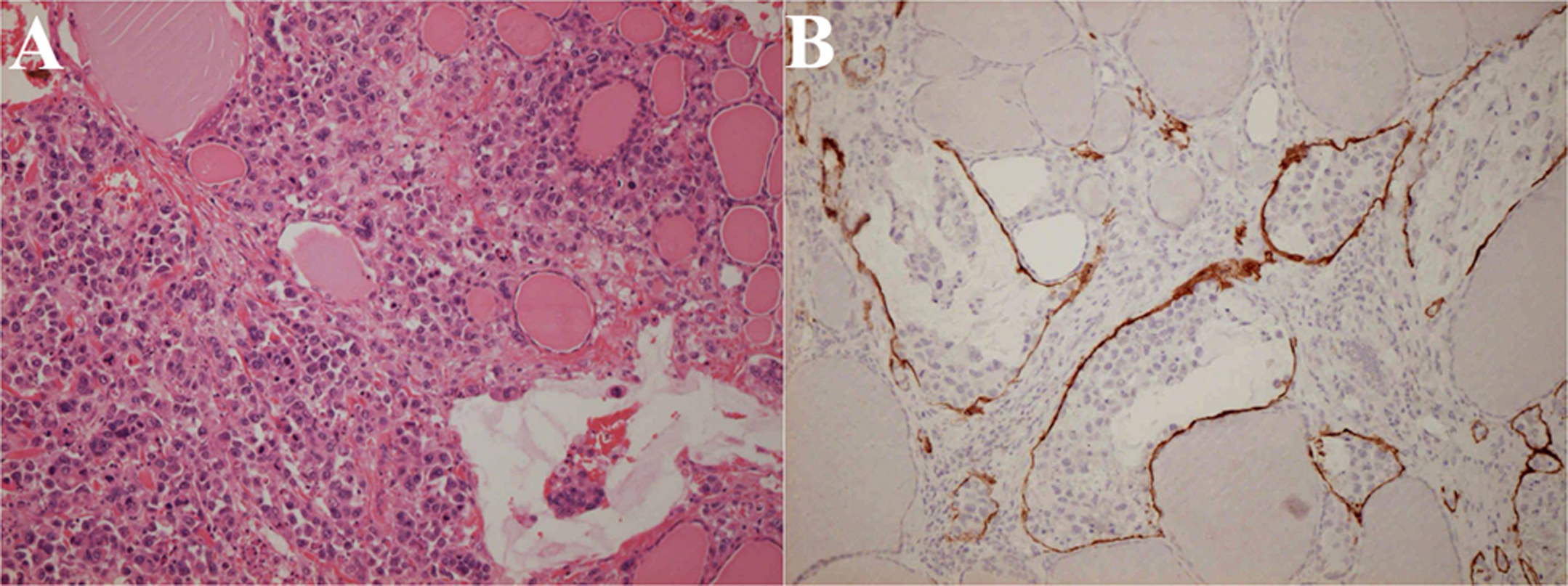
Figure 4. Histopathology of the thyroid gland at autopsy. The thyroid gland is destructed by extensive tumor invasion and colloid content is diminished (A: Hematoxylin and eosin stain; × 200). Lymph vessels are filled with tumor cells (B: Immunohistochemical stain using D2-40 monoclonal antibodies that specifically stain lymph vessels; × 200).