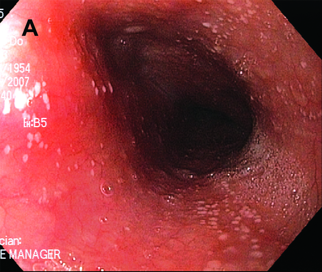
Figure A. Esophagoscopy revealed numerous 1-2 mm white plaques that was adherent from mid-esophagus down to the gastroesophageal junction.
| Gastroenterology Research, ISSN 1918-2805 print, 1918-2813 online, Open Access |
| Article copyright, the authors; Journal compilation copyright, Gastroenterol Res and Elmer Press Inc |
| Journal website http://www.gastrores.org |
Case Report
Volume 3, Number 4, August 2010, pages 173-174
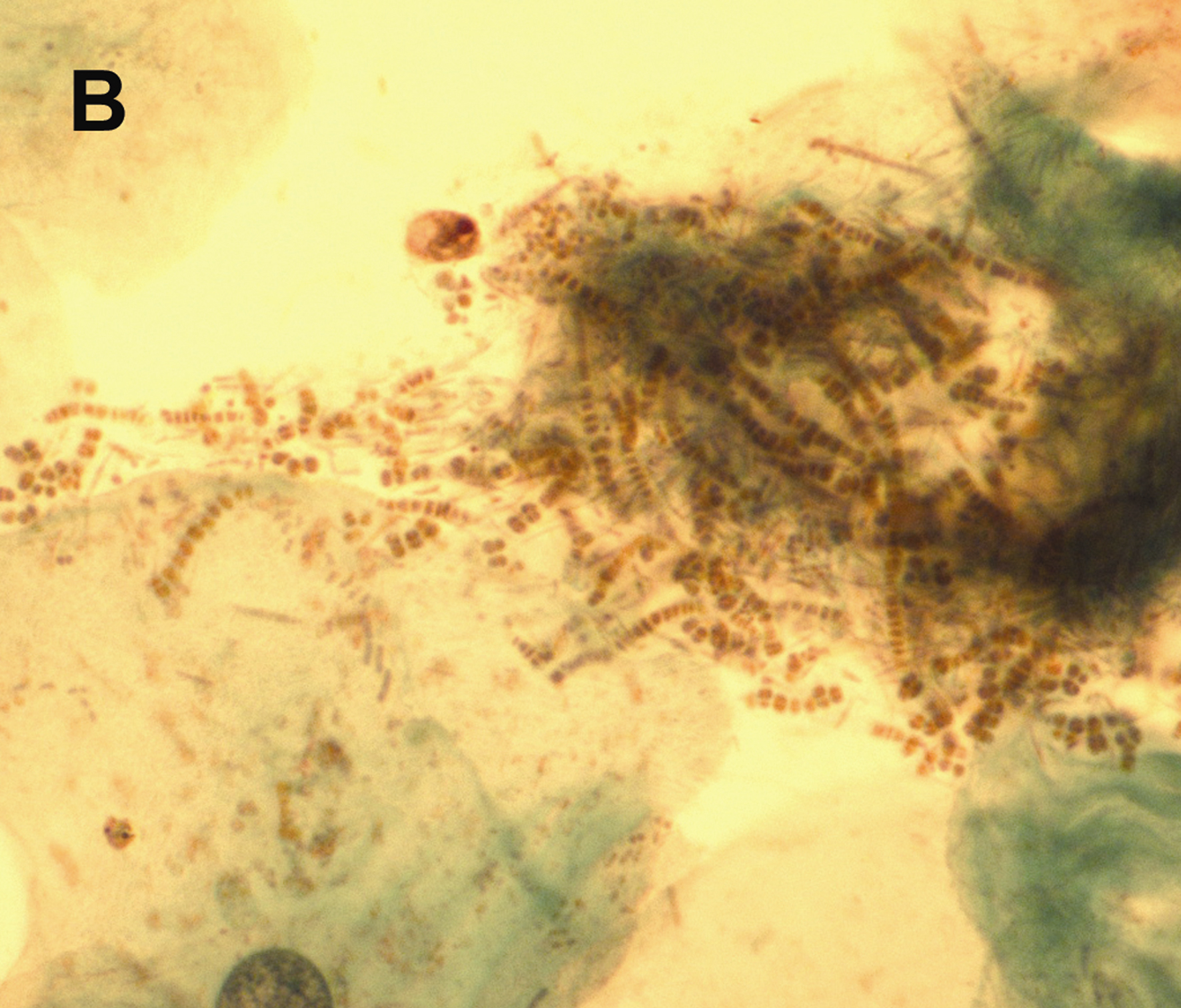
Dermatophilus Congolensis Infection of the Esophagus
Figures