| 1 | 68 | F | Malaise, weight loss, anemia | 11 | + | Chemotherapy; partial hepatectomy | [11] |
| 2 | 35 | F | Epigastric discomfort, fever, weight loss | 20 | + | Right hemihepatectomy | [5] |
| 3 | 37 | M | Malaise, weight loss | 15 | + | Right trisegmentectomy with caudate lobectomy | [16] |
| 4 | 19 | F | R't upper quadrant pain, weight loss, palpable abdominal mass | 12 | + | Excision | [18] |
| 5 | 56 | F | Gastrointestinal upset | 15 | + | Resection | [18] |
| 6 | 40 | F | Epigastric pain, weight loss | 12.5 | + | Left hepatectomy | [18] |
| 7 | 49 | F | Liver mass on ultrasound during routine health check-up | 4.2 | + | Excision | [18] |
| 8 | 31 | F | Abdominal distention, weight loss | 15 | + | Right hemihepatectomy | [18] |
| 9 | 57 | F | Epigastralgia | 9.5 | + | Right hemihepatectomy | [17] |
| 10 | 51 | F | Abdominal discomfort, weight loss | 12 | + | Left lobectomy | [18] |
| 11 | 30 | F | Liver mass on ultrasound during routine health check-up | 5.5 | + | Right lobectomy | [19] |
| 12 | 82 | M | Liver mass on abdominal CT scan during evaluation of renal calculi; weight loss, weakness | 10 | - | Right lobectomy | [21] |
| 13 | 57 | F | Abdominal pain, dizziness, vomiting | NA | + | Resection | [4] |
| *14 | 70 | M | Abdominal pain, weight loss | 2.5 | - | NA | [6] |
| *15 | 65 | F | Staging workup showed lesion in liver from abdominal CT | 2 | NA | Resection | [7] |
| *16 | 19 | M | Right upper quadrant pain | 16 | NA | Chemotherapy# | [8] |
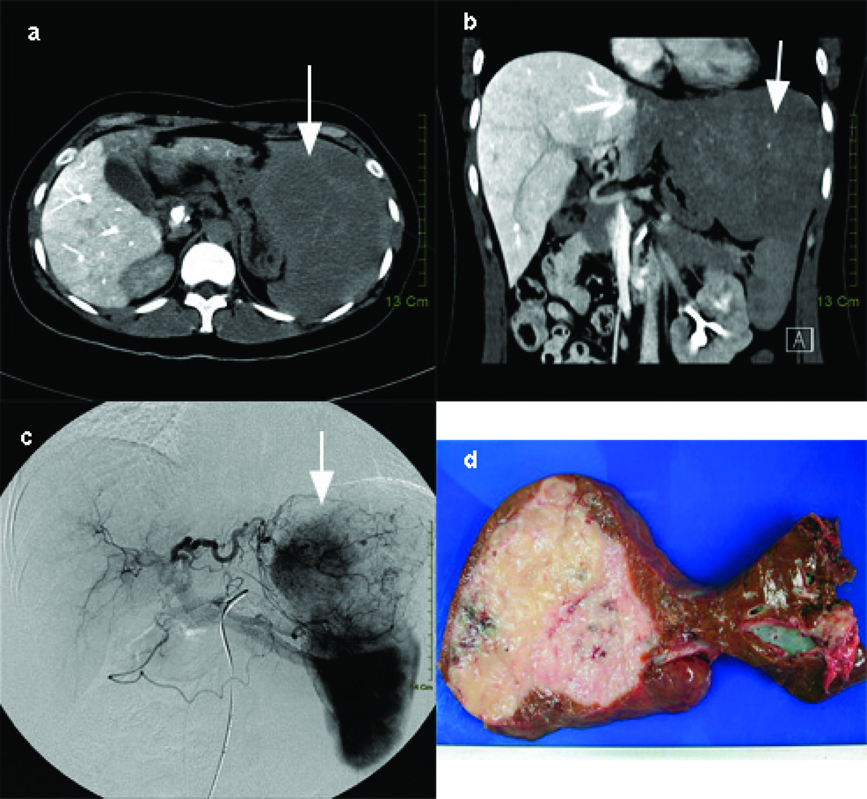
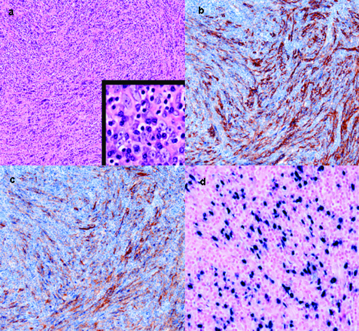
| 17 | 36 | F | Epigastric discomfort | 14 | + | Left Lateral segmental hepatectomy | Present case |