Gastroenterology Research, ISSN 1918-2805 print, 1918-2813 online, Open Access
Article copyright, the authors; Journal compilation copyright, Gastroenterol Res and Elmer Press Inc
Journal website http://www.gastrores.org

Original Article

Volume 3, Number 2, April 2010, pages 79-85


Traumatic Transection of Pancreas at the Neck: Feasibility of Parenchymal Preserving Strategy

Figures

Figure 1.
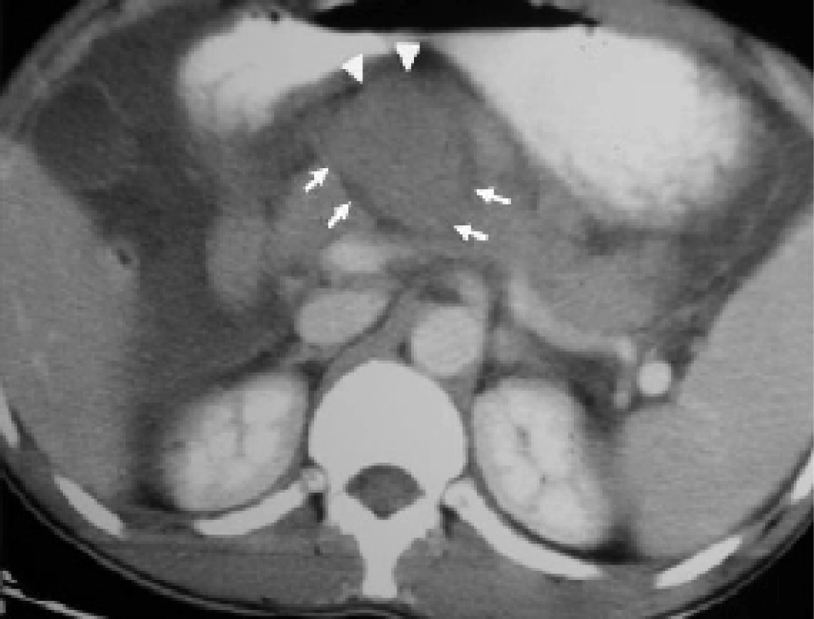
Figure 1. CECT abdomen axial image reveals complete transection between head and neck of pancreas with a rounded hypoechoic mass suggestive of hematoma separating the two fractured fragments (arrows). Note the fluid surrounding the hematoma (arrow heads).
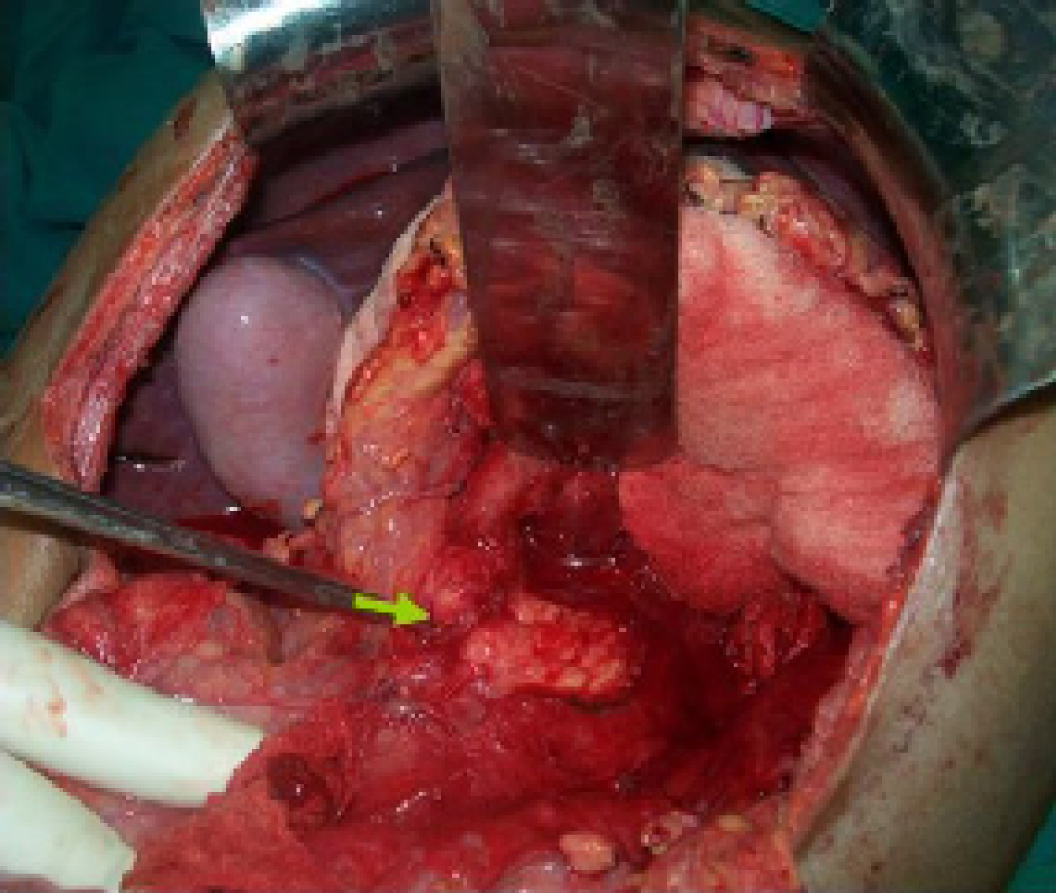
Figure 2.
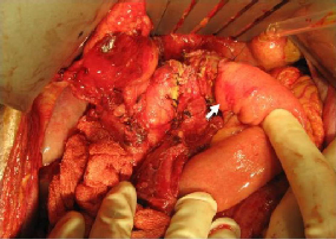
Figure 2. Intraoperative photograph shows the pancreatic fracture (arrow)
Figure 3.
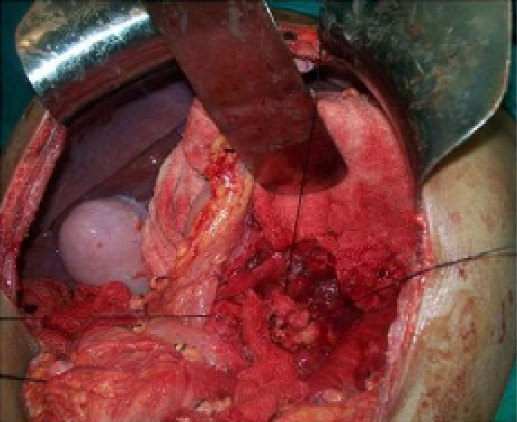
Figure 3. The distal pancreas is lifted for a distance of 2 - 3 cm.
Figure 4.
Figure 4. Anastomosis of the distal pancreas to Roux-en-Y jejunal loop.
Figure 5.
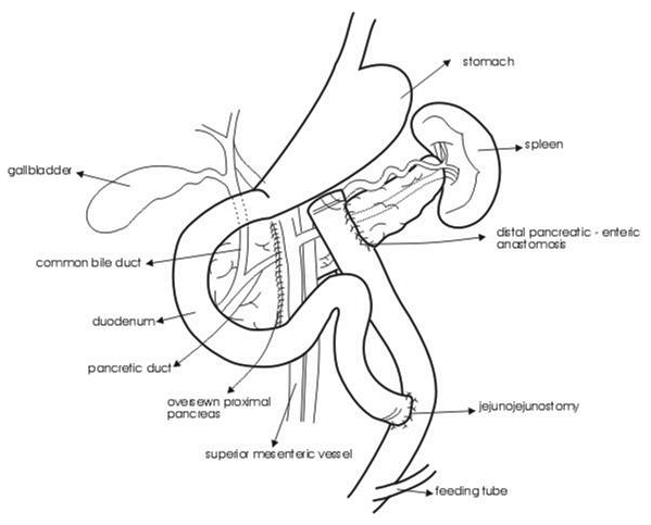
Figure 5. Line diagram describing the operative details.

Table

Table 1. Patients Demographic Characteristics
 
ParameterCase 1Case 2
Age (years)/Sex15/Male20/Male
Duration2 weeks24 hours
MechanismBicycle handle-barBicycle handle-bar
Clinical presentationAscites, dehydration, Tense abdomenEpigastric tenderness
CT abdomenAscites, Transection neckTransection neck
Operative findingsAscites, complete transectionHematoma complete transection
Operative procedurePancreaticojejunostomyPancreaticojejunostomy
OutcomeRecoveredRecovered