Gastroenterology Research, ISSN 1918-2805 print, 1918-2813 online, Open Access
Article copyright, the authors; Journal compilation copyright, Gastroenterol Res and Elmer Press Inc
Journal website http://www.gastrores.org

Review

Volume 3, Number 1, February 2010, pages 1-8


Hepatobiliary Manifestations of Sickle Cell Anemia

Figures

Figure 1.
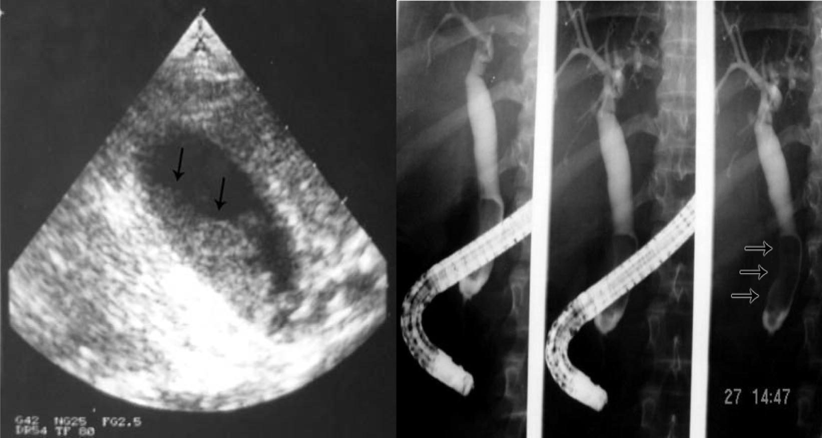
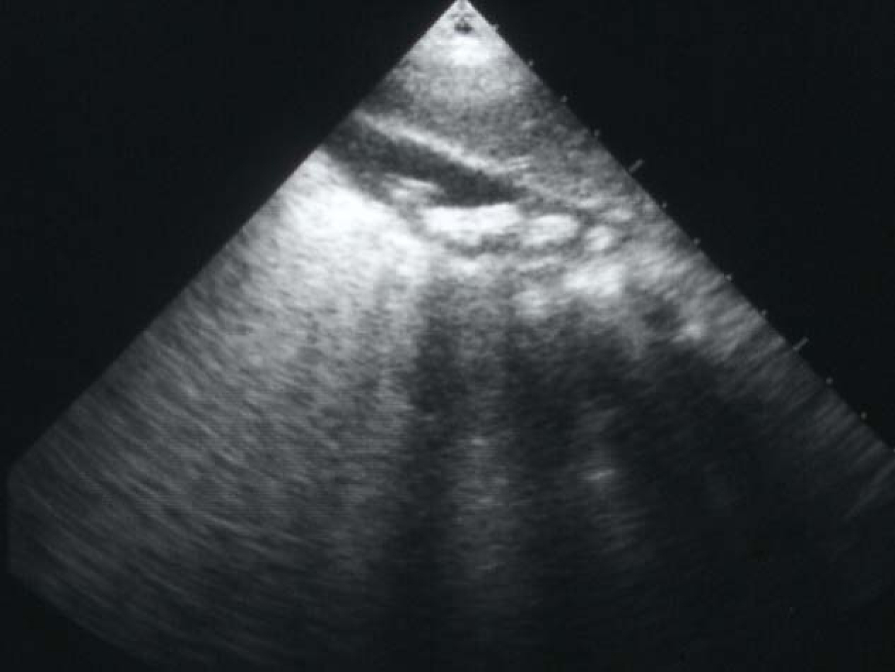
Figure 1. Abdominal ultrasound showing multiple gallstones.
Figure 2.
Figure 2. Intraoperative photograph showing acute gangrenous cholecystitis in a patient with SCA.
Figure 3.
Figure 3. Percutaneous cholangiogram showing gallstones and common bile duct stones.
Figure 4.
Figure 4. ERCP showing bile duct stones pre and post laparoscopic cholecystectomy.
Figure 5.
Figure 5. Biliary sludge in the gallbladder and bile ducts.
Figure 6.
Figure 6. ERCP showing dilated bile ducts without an obstructive cause (Sickle cell cholangiopathy).
Figure 7.
Figure 7. CT-scan of the abdomen showing the liver with iron overload in a patient with SCA and multiple blood transfusions.