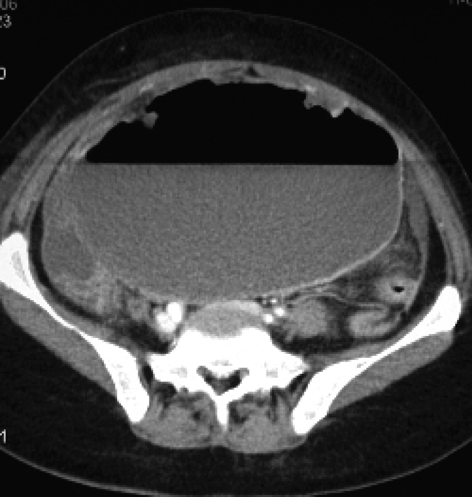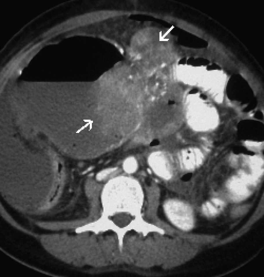
Figure 1. A postcontrast computed tomographic scan showed a huge primary calcified GIST (arrows) with a cystic component.
| Gastroenterology Research, ISSN 1918-2805 print, 1918-2813 online, Open Access |
| Article copyright, the authors; Journal compilation copyright, Gastroenterol Res and Elmer Press Inc |
| Journal website http://www.gastrores.org |
Case Report
Volume 2, Number 6, December 2009, pages 361-363
Spontaneously Ruptured Gastrointestinal Stromal Tumor With Pelvic Abscess: A Case Report and Review
Figures