Gastroenterology Research, ISSN 1918-2805 print, 1918-2813 online, Open Access
Article copyright, the authors; Journal compilation copyright, Gastroenterol Res and Elmer Press Inc
Journal website http://www.gastrores.org

Case Report

Volume 2, Number 5, October 2009, pages 303-306


Obstructive Jaundice Due to Hilar Bile Duct Compression with Encasement of the Right Hepatic Artery

Figures

Figure 1.
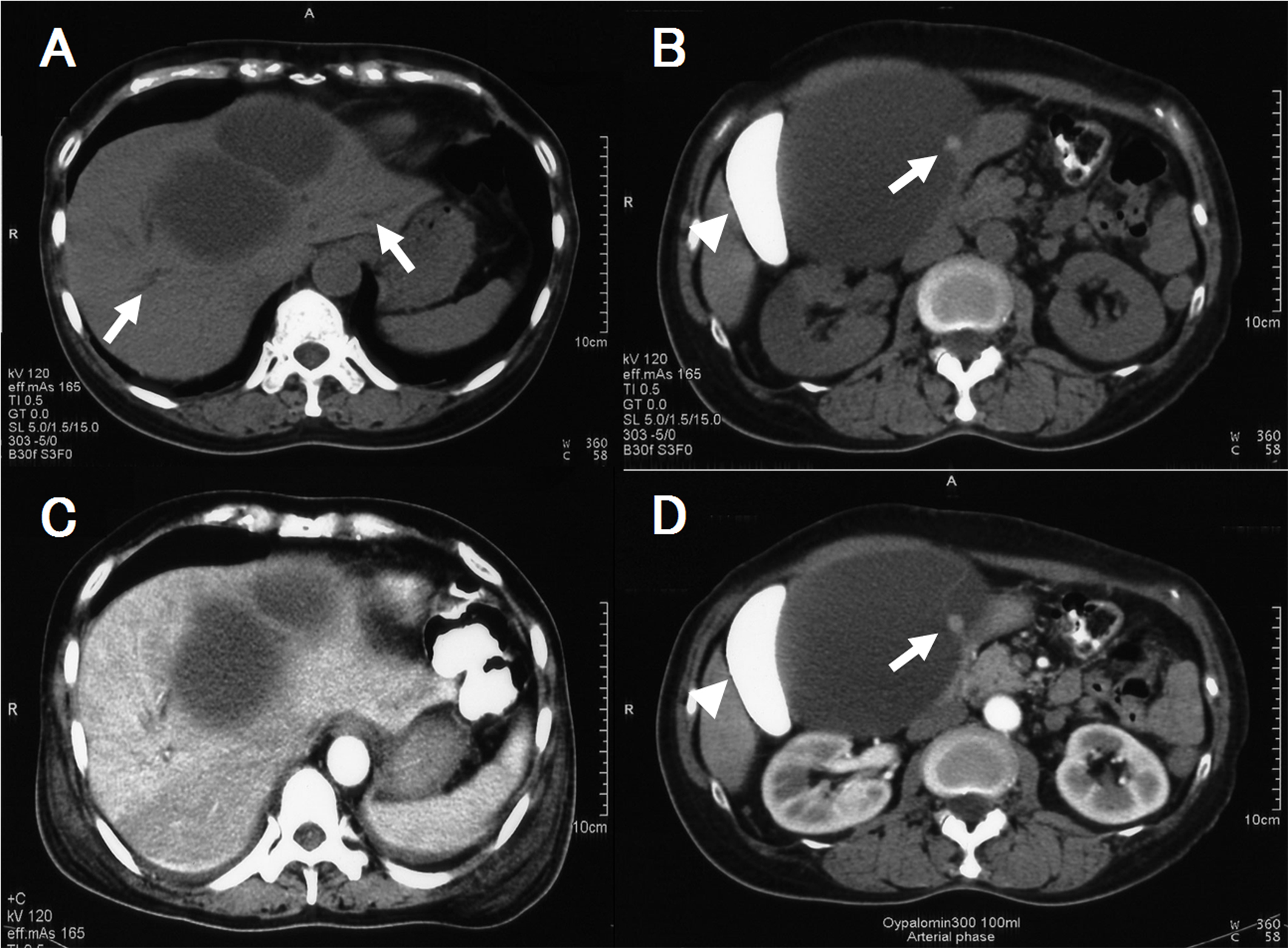
Figure 1. A and B: Plain CT after drip infusion CT cholangiography (DIC-CT), C and D: Enhanced CT after DIC-CT. A and C showed a hepatic cyst with septum near the hepatic hilum and slight dilatation of the intrahepatic duct (arrow). B and D showed a mural module which did not enhance with contrast material (arrow) as well as deformation of the gallbladder (arrowhead).
Figure 2.
Figure 2. A: Endoscopic retrograde cholangiography (ERC) demonstrated a band-like filling defect of the hilar bile duct (arrow). B: Choledochoscopy demonstrated compression and mucosal erosive changes of the hilar bile duct.
Figure 3.
Figure 3. A: Abdominal angiography revealed distortion of the cystic and gastroduodenal arteries as well as slight encasement of the right hepatic artery (arrow). B: Mechanical displacement of the portal vein was observed, and the left branch of the portal vein was not visualized.
Figure 4.
Figure 4. A: The filling defect of the bile duct disappeared on ERCP. B: Choledochoscopicexamination revealed normal bile duct without stenosis nor mucosal erosive changes.