Gastroenterology Research, ISSN 1918-2805 print, 1918-2813 online, Open Access
Article copyright, the authors; Journal compilation copyright, Gastroenterol Res and Elmer Press Inc
Journal website http://www.gastrores.org

Case Report

Volume 2, Number 2, April 2009, pages 118-121


Esophageal Carcinoma with Triplicate Differentiation into Squamous Cell Carcinoma, Small Cell Carcinoma and Adenocarcinoma: a Case Report

Figures

Figure 1.
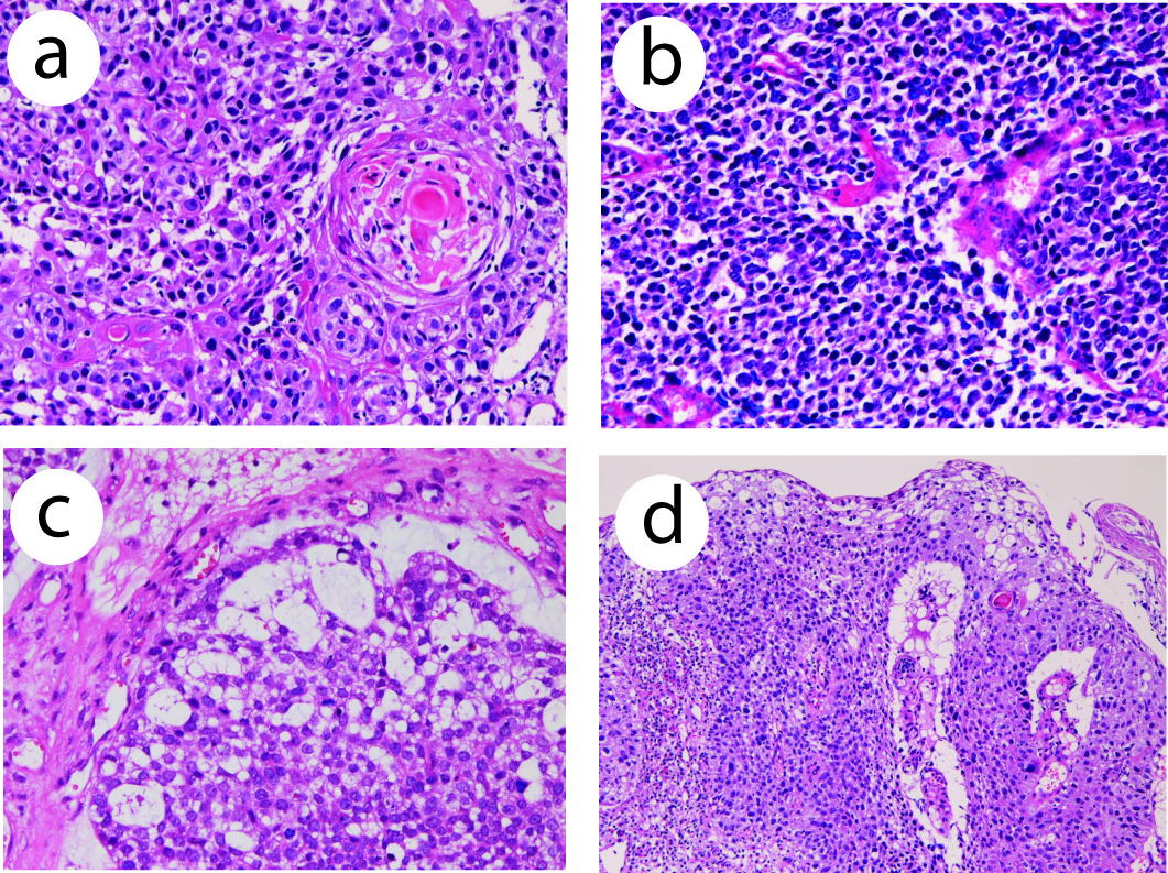
Figure 1. (a) Histology of squamous cell carcinoma element of the esophageal tumor. Keratinization is seen. HE, x 200. (b) Small cell carcinoma element of the esophageal carcinoma. The tumor cells shows characteristic morphologies of small cell carcinoma. HE, x 200. (c) Adenocarcinoma element of the esophageal carcinoma. Tubular formations are seen. There is a gradual transition between adenocarcinoma element and small cell carcinoma element. HE, x 200. (d) Transitional zone between squamous cell carcinoma element and small cell carcinoma element of the esophageal carcinoma. HE, x 100.
Figure 2.
Figure 2. (a) High molecular weight cytokeratin is positive in the squamous cell carcinoma element. Immunostaining (AE1/3), x 200. (b) p53 expression in squamous cell carcinoma element of the esophageal tumor. Immunostaining, x 200. (c) Synaptophysin is positive in the small cell carcinoma element of the esophageal tumor cells. Immunostaining, x 200. (d) Ki-67 labeling in the small cell carcinoma element of the esophageal tumor. The labeling is 95%. Immunostaining, x 200.