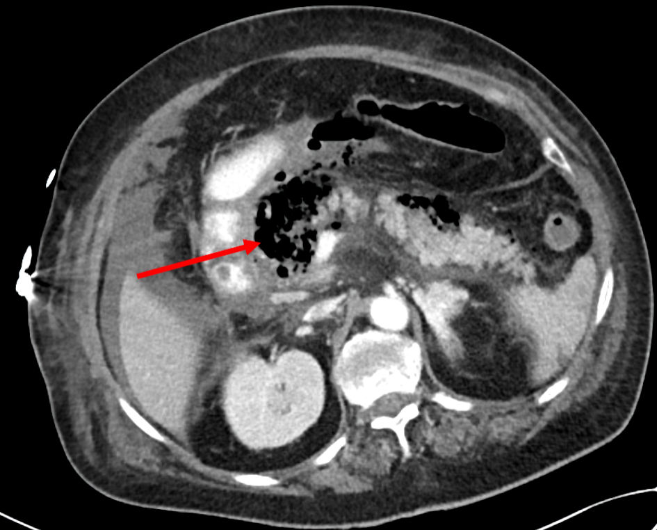
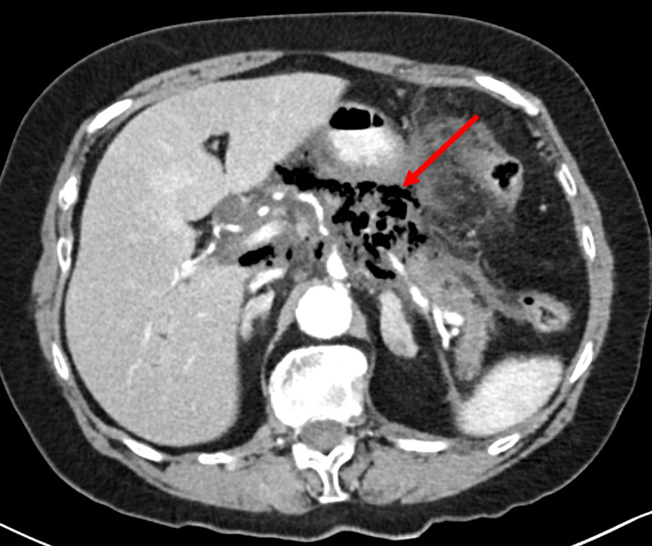
Figure 1. An axial abdomino-pelvic CT scan showed multilocular pancreatic collection involving the lesser sac containing air (arrow), consistent with acute emphysematous pancreatitis. CT: computed tomography.
| Gastroenterology Research, ISSN 1918-2805 print, 1918-2813 online, Open Access |
| Article copyright, the authors; Journal compilation copyright, Gastroenterol Res and Elmer Press Inc |
| Journal website https://www.gastrores.org |
Case Report
Volume 17, Number 1, February 2024, pages 32-36
Fulminant Emphysematous Pancreatitis: Diagnosis Time Counts
Figures
Table
| Case 1 | Case 2 | |
|---|---|---|
| CT: computed tomography; ICU: intensive care unit; IV: intravenous; COPD: chronic obstructive pulmonary disease; NASH: non-alcoholic steatohepatitis. | ||
| Patient’s age, years | 78 | 71 |
| Sex | Female | Female |
| Medical history | Hypertension, hyperlipidemia | COPD, NASH, aortic stenosis |
| Clinical presentation | Abdominal pain and vomiting | Abdominal pain and vomiting |
| Diagnosis time (by CT scan) | 52 h | 7 h |
| ICU admission and close monitoring time | 48 h | 8 h |
| IV antibiotics | 48 h | 7 h |
| Outcome | Died | Survived |