Gastroenterology Research, ISSN 1918-2805 print, 1918-2813 online, Open Access
Article copyright, the authors; Journal compilation copyright, Gastroenterol Res and Elmer Press Inc
Journal website https://www.gastrores.org

Review

Volume 16, Number 5, October 2023, pages 249-253


Gastrointestinal Mucormycosis: A Clinical Review

Figures

Figure 1.
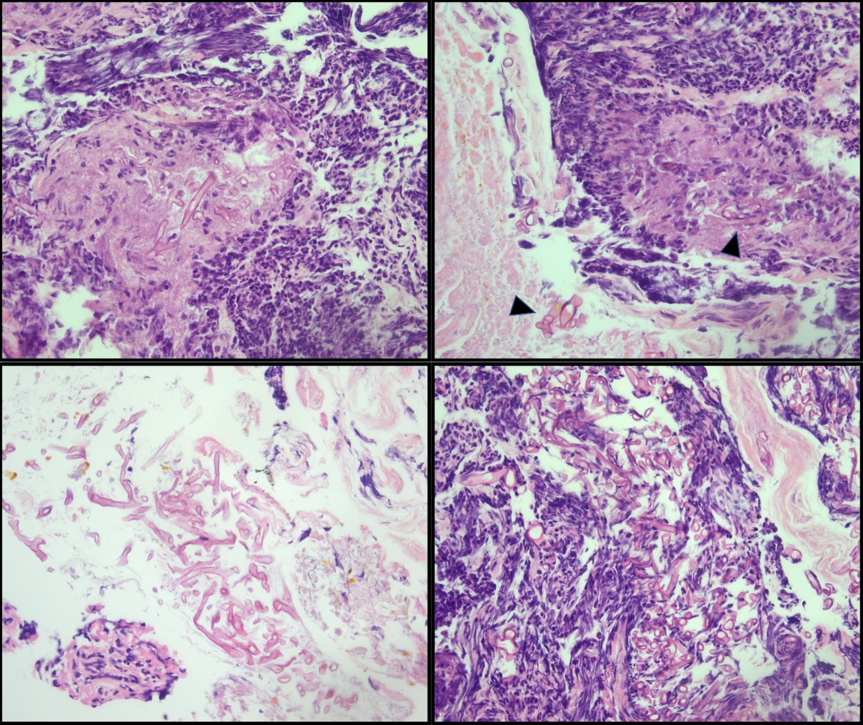
Figure 1. Cecal biopsy. Large, non-septate fungal organisms consistent with Mucorales (Mucor/Rhizopus) in a background of ulceration and acute inflammation (arrowhead). The presence of fungal organisms in the ulcer bed is most likely indicative of infection.
Figure 2.
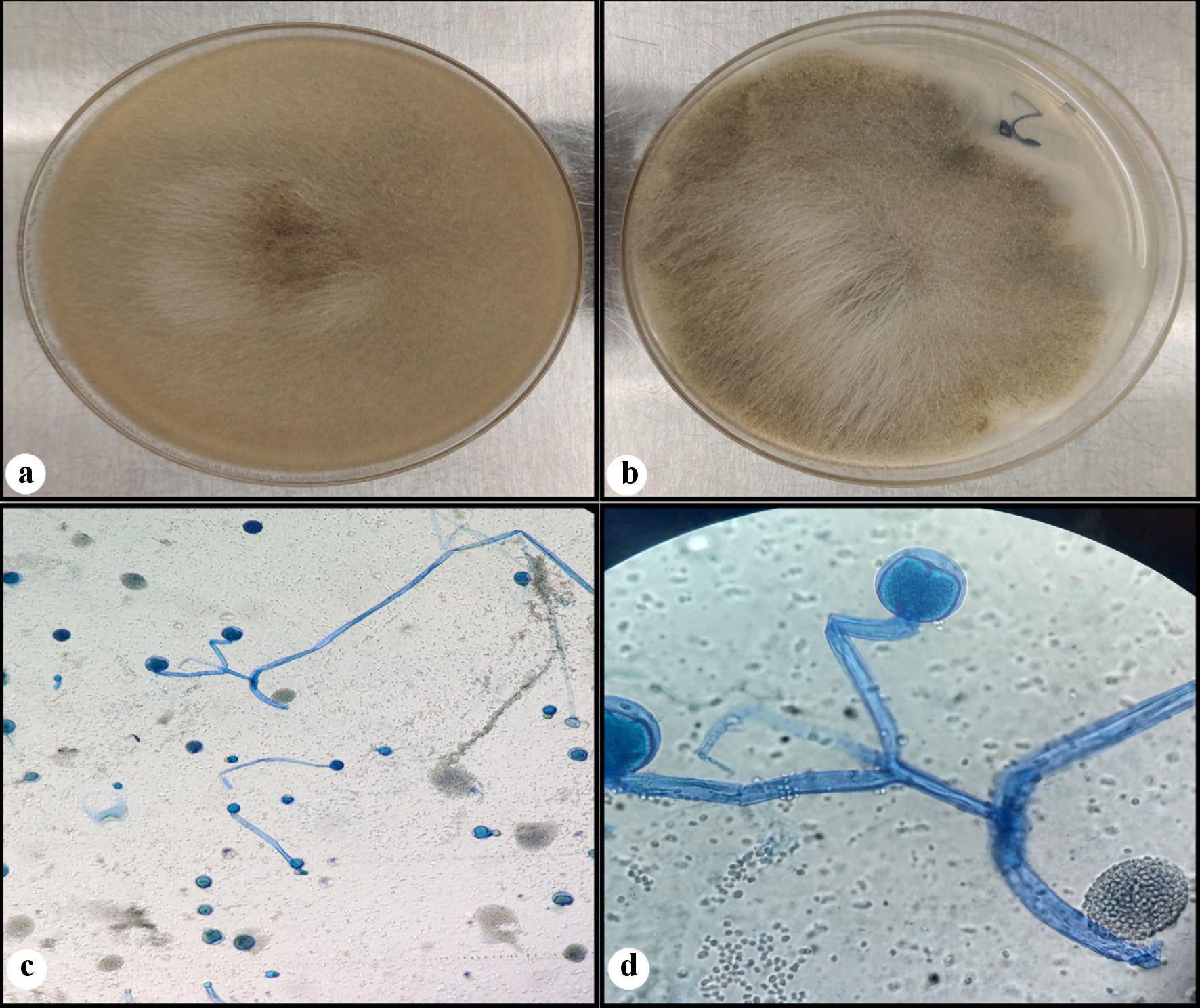
Figure 2. Fungal culture. Rapidly growing white fluffy colony that overfills agar plate on the Sabouraud dextrose agar (SDA) (a), and almost completely filling the potato dextrose agar (PDA) (b). A lactophenol cotton blue (LCB) preparation (c, d) showing wide hyphae, rare septations, branched sporangiophores, with round sporangia (sac-like structure). (d) Seen in the background are some spores of ruptured sporangium.

Table

Table 1. Primary and Combination Antifungal Therapy for the Treatment of Systemic Mucormycosis Including Gastrointestinal Involvement
 
Primary antifungal therapyRecommended dosageComments
CNS: central nervous system.
Liposomal amphotericin B5 - 10 mg/kg/dayExpensive, less nephrotoxic, better CNS penetration than other formulations.
Amphotericin B deoxycholate1.0 - 1.5 mg/kg/dayHighly toxic, inexpensive, only licensed agent.
Amphotericin B lipid complex5 - 7.5 mg/kg/dayMore nephrotoxic than liposomal amphotericin B, murine data suggest possible benefits in combination with echinocandins.