| Age (years) | 75 | 48 | 44 | 22 |
| Gender | Male | Female | Male | Male |
| Past medical history | Atrial fibrillation, chronic hepatitis B, liver cirrhosis, and HCC | Inflammatory cecal mass and recurrent multiple liver abscesses | None | None |
| Cirrhosis (Child-Pugh score) | Yes (10) | No | No | No |
| History of chronic hepatitis | Hepatitis B | No | No | No |
| Previous history of liver abscess | None | Yes | No | No |
| Presentation | Fever, epigastric pain, abdominal distention, anorexia, jaundice | Fever, epigastric pain, abdominal distention | Fever, chills, right upper abdominal pain and shortness of breath | Shortness of breath, right-sided chest pain and RUQ abdominal pain |
| Initial CRP (mg/L) | 249 | 240 | 201 | 250 |
| Alkaline phosphatase | 70 | 46 | 186 | 332 |
| Albumin | 3.1 | 2.8 | 2.9 | 3.7 |
| Aspirate culture (intraoperative cultures) | Enterococcus faecium (E. coli) | No growth | No growth | Pseudomonas aeruginosa |
| Blood cultures | Negative | Negative | Negative | Negative |
| Source of the abscess | | | Unknown | Traumatic (shrapnel injury after explosion) |
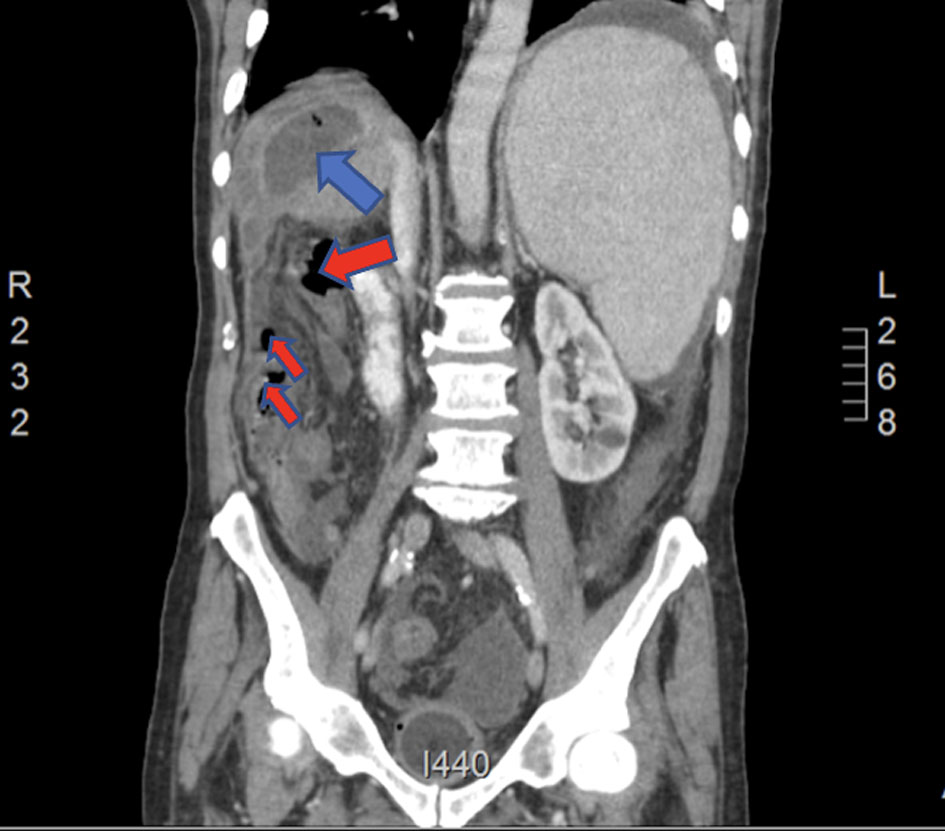
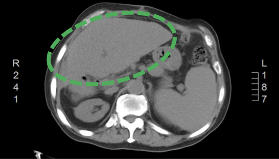
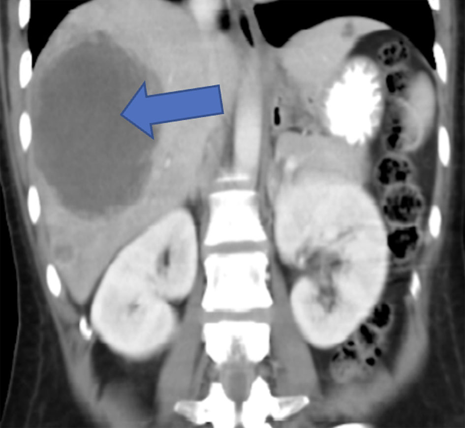
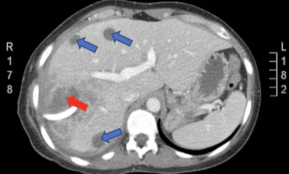
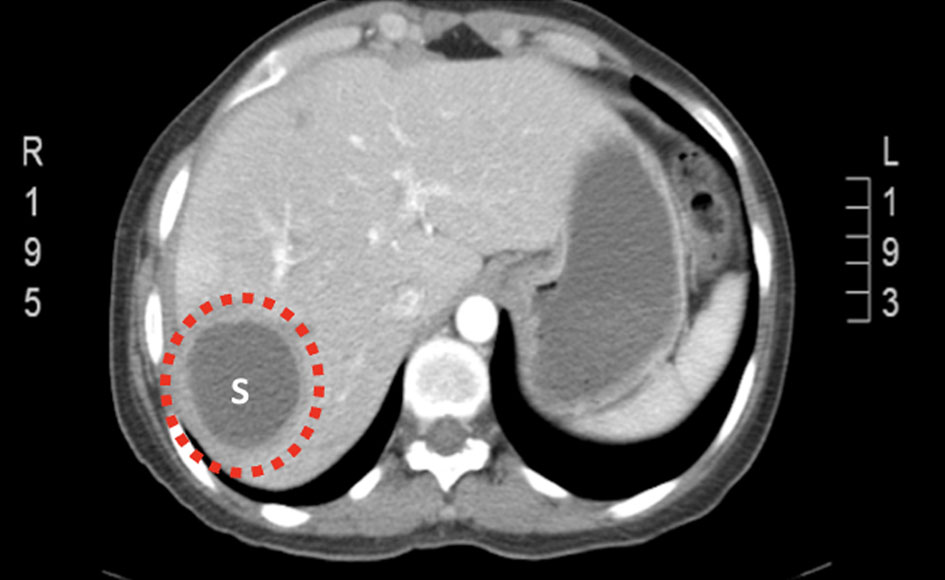
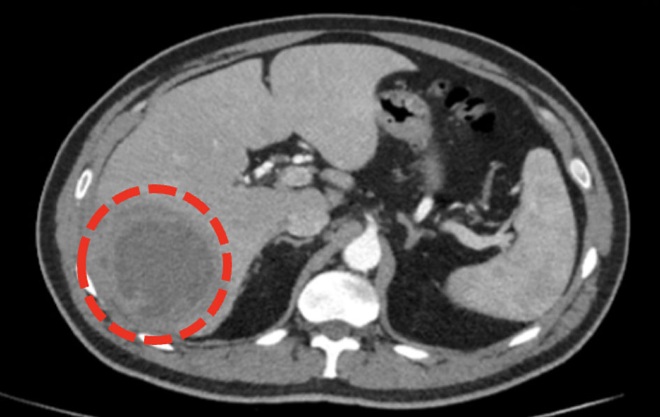
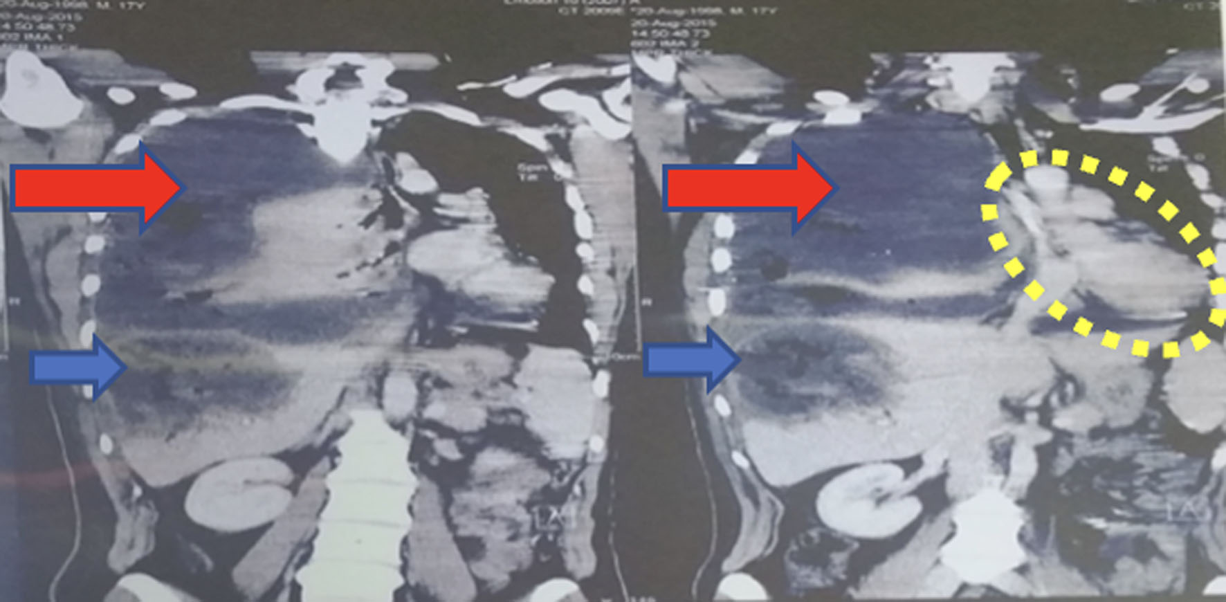
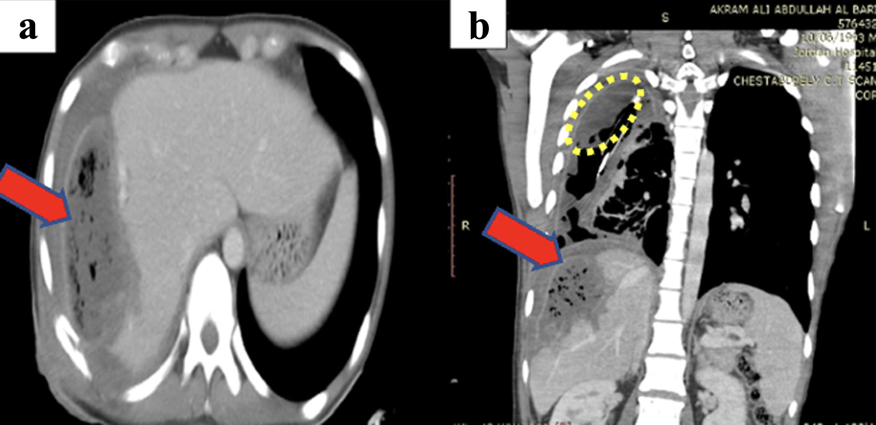
| Liver abscess site and location | Segment VII, measuring (7 × 5 cm) | Segment VII, multiple abscesses largest (11.6 × 10.4 cm) | Segment VII (5 × 6.1 cm) cm and segment VI (8.7 × 8 cm) | Lateral aspect of the right hepatic lobe, approximately 11 × 10 × 7 cm |
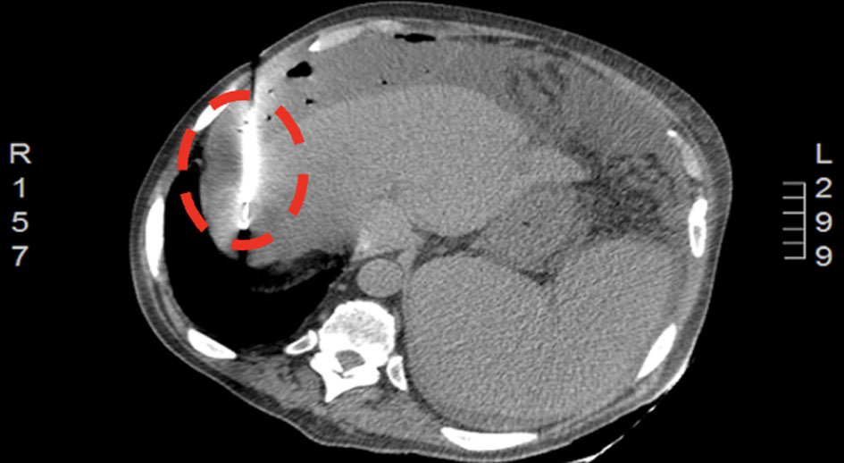
| Percutaneous drain trial (modality, number) | Yes (CT-guided aspiration, once) | Yes (CT-guided drainage, twice) | No | Yes (ultrasound-guided drainage, once) |
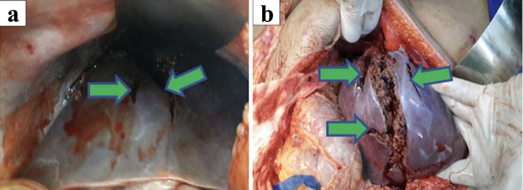
| Surgical modality | Open drainage with resection of segment VII | Cholecystectomy with resection of segment VII and most of segment VI | Cholecystectomy with resection of segment VII and segment VI | Right hepatectomy |
| A follow-up modality and timing | CT of abdomen and pelvis 3 months later | Ultrasound 3 weeks later | Ultrasound after 1 week | Ultrasound 3 weeks |
| Follow-up condition | Improved | Improved | Improved | Improved |
| Follow-up CRP (mg/dL) | 39 | 146 | 147 | 86 |