Gastroenterology Research, ISSN 1918-2805 print, 1918-2813 online, Open Access
Article copyright, the authors; Journal compilation copyright, Gastroenterol Res and Elmer Press Inc
Journal website https://www.gastrores.org

Case Report

Volume 16, Number 2, April 2023, pages 118-124


Esophageal Ulcer After Intravitreal Ranibizumab Injection in a Patient With Age-Related Macular Degeneration

Figures

Figure 1.
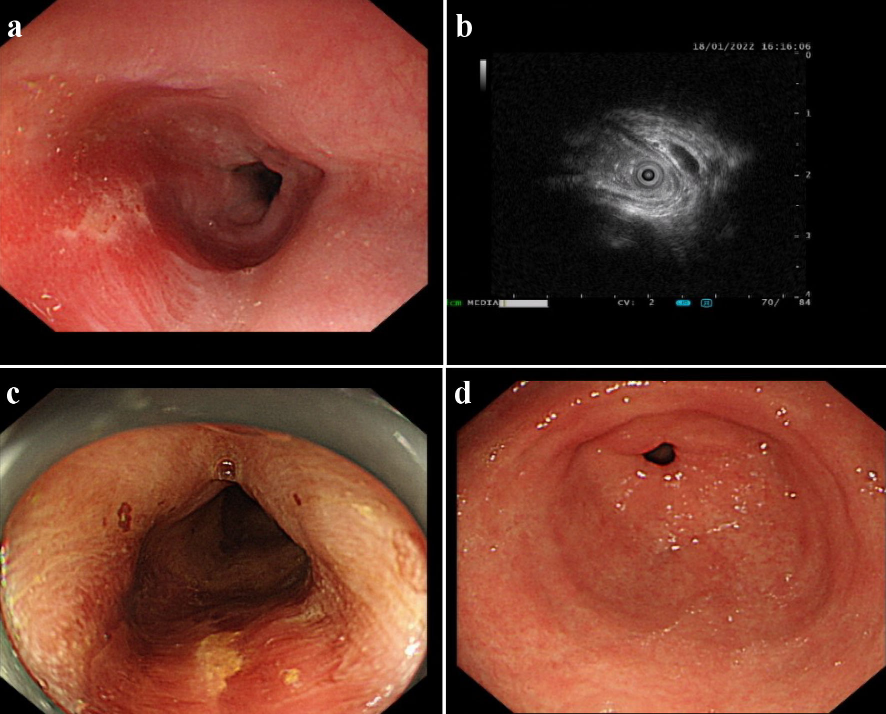
Figure 1. An esophageal surface ulcer occurred after intravitreal ranibizumab injection. (a) Ultrasound gastroscopy showed an esophageal surface ulcer 20 - 24 cm from the incisor. The central of ulcer was covered with fibrinous tissue, and the surrounding mucosae were flushing and congestive. (b) Ultrasonography revealed that the mucosae were slightly incrassated with a hypoechoic variation in the lesion area. (c) The fibrinous tissue was stained by Lugol solution. (d) Ultrasound gastroscopy showed that the mucosae in the gastric antrum were smooth.
Figure 2.
Figure 2. The boundary of the esophageal ulcer was visualized using magnifying endoscopy with narrow-band imaging (ME-NBI). ME-NBI showed the boundary of the esophageal ulcer with color transformation to hazel, and no abnormal blood vessel was observed, indicating that neither intestinal metaplasia nor cancerization occurred in the esophageal ulcer.
Figure 3.
Figure 3. The esophageal biopsy revealed the pathological characteristic of the lesion using the optical microscope.
Figure 4.
Figure 4. Timeline of ranibizumab-induced esophageal ulcer in a patient with age-related macular degeneration. AMD: age-related macular degeneration; PPI: proton pump inhibitor; TCM: traditional Chinese medicine.