Gastroenterology Research, ISSN 1918-2805 print, 1918-2813 online, Open Access
Article copyright, the authors; Journal compilation copyright, Gastroenterol Res and Elmer Press Inc
Journal website https://www.gastrores.org

Original Article

Volume 16, Number 1, February 2023, pages 9-16


Endoscopic Ultrasound Predicts Risk of Occult Intra-Abdominal Metastases in Localized Gastric Cancer: A Validation Study

Figures

Figure 1.
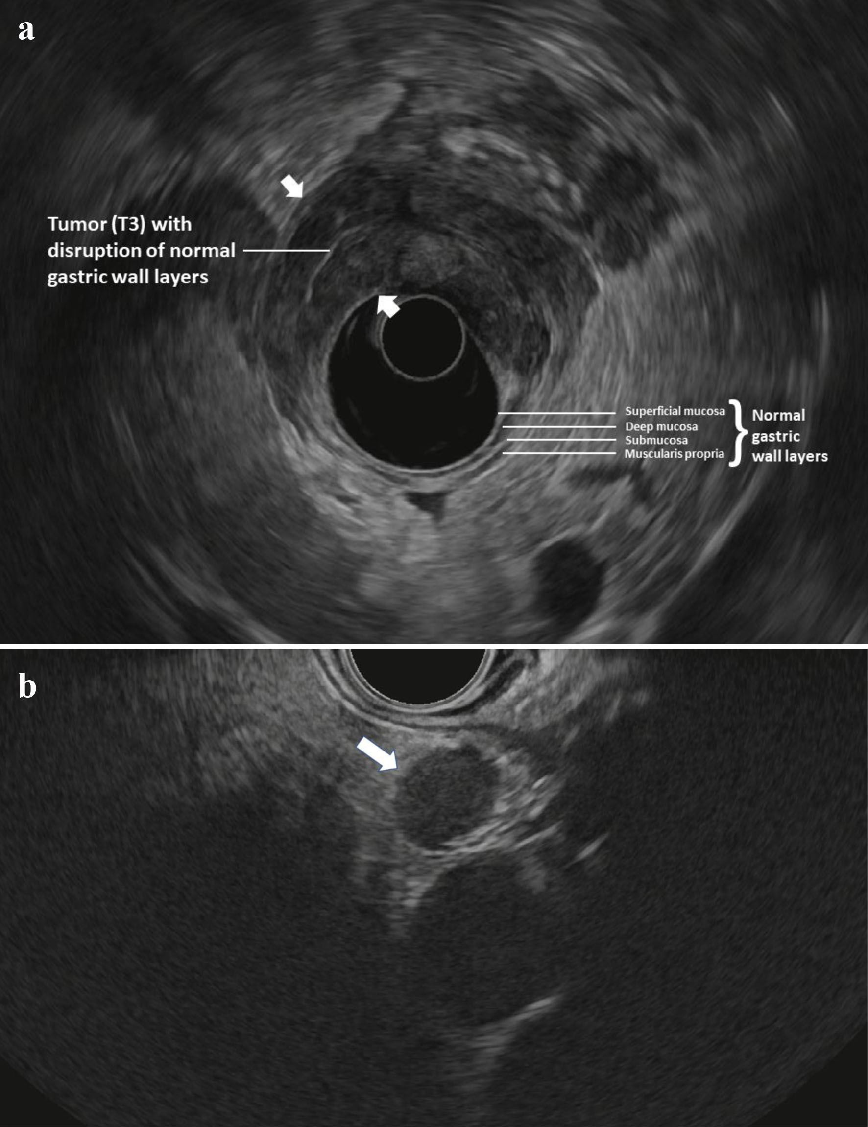
Figure 1. (a) Radial EUS image of a T3 antral tumor showing normal gastric layers on EUS and the large tumor (between arrowheads) disrupting the normal layers and invading through the muscularis propria and into the serosa. This patient was classified as “high-risk” and ultimately was found to have M1 disease on DSL. (b) Linear EUS image of an N+ tumor showing a malignant appearing lymph node (arrow). EUS: endoscopic ultrasound; DSL: diagnostic staging laparoscopy.
Figure 2.
Figure 2. Cytospin from peritoneal fluid shows a cluster of tumor cells with focal signet ring cell differentiation (arrow) (Papinocolou stain, × 600).

Tables

Table 1. Patient and Tumor Characteristics
 
CharacteristicOverall (n = 68)Laparoscopy M0 (n = 51)Laparoscopy M1 (n = 17)P-value
GEJ: gastroesophageal junction.
Gender0.773
  Male42 (62%)31 (61%)11 (65%)
  Female26 (38%)20 (39%)6 (35%)
Age (years)
  Median (range)64 (16 - 87)65 (16 - 87)64 (45 - 80)0.639
  ≥ 6047 (69%)36 (71%)11 (65%)0.649
Ethnicity/race0.370
  Caucasian (White)34 (50%)26 (51%)8 (47%)
  African American19 (28%)16 (31%)3 (18%)
  Hispanic8 (12%)4 (8%)4 (24%)
  Asian6 (9%)4 (8%)2 (12%)
  Native American1 (1%)1 (2%)0
Tumor location0.088
  Body25 (37%)20 (39%)5 (29%)
  Antrum21 (31%)16 (31%)5 (29%)
  Whole stomach13 (19%)6 (12%)7 (42%)
  Cardia6 (9%)6 (12%)0
  GEJ3 (4%)3 (6%)0
Histology
  Adenocarcinoma67 (99%)50 (98%)17 (100%)
  Laurens classification0.495
    Diffuse28 (41%)20 (39%)8 (47%)
    Intestinal17 (25%)15 (29%)2 (12%)
    Not reported19 (28%)13 (25%)6 (35%)
    Mixed4 (6%)3 (6%)1 (6%)
Histologic differentiation0.317
  G1: Well-differentiated1 (1%)1 (2%)0
  G2: Moderately-differentiated8 (12%)8 (16%)0
  G3: Poorly-differentiated53 (78%)38 (74%)15 (88%)
  Not reported6 (9%)4 (8%)2 (12%)
Signet ring cell morphology27 (40%)19 (37%)8 (47%)0.474


Table 2. Endoscopic Characteristics
 
CharacteristicOverallLaparoscopy M0Laparoscopy M1P-value
*Significant (P < 0.05).
Linitis plastica appearance17 (25%)9 (18%)8 (47%)0.015*
Ulceration47 (69%)36 (71%)11 (65%)0.649
Circumferential involvement28 (41%)17 (33%)11 (65%)0.023*
EUS T stage0.494
  T11 (1%)1 (2%)0
  T25 (7%)5 (10%)0
  T359 (87%)42 (82%)17 (100%)
  T43 (4%)3 (6%)0
EUS N stage0.024*
  N020 (29%)18 (36%)2 (12%)
  N125 (37%)15 (29%)10 (59%)
  N217 (25%)15 (29%)2 (12%)
  N36 (9%)3 (6%)3 (18%)
EUS M stage1
  M068 (100%)51 (100%)17 (100%)
EUS ascites2 (3%)2 (4%)01


Table 3. Diagnostic Laparoscopy Characteristics
 
CharacteristicN%
Peritoneal washing
  Negative5175
  Positive1624
  Not done11
Positive cytology
  Metastatic adenocarcinoma1169
  Atypical cells531
Visible metastatic disease
  No6291
  Yes69
Location of visible disease
  Peritoneum466%
  Omentum117%
  Diaphragmatic caking117%
Ascites
  No6190%
  Yes710%
Overall laparoscopic M stage
  M05175%
  M11725%


Table 4. Endoscopic Ultrasound High-Risk and Low-Risk Subdivision and Laparoscopic Identification of Occult M1 Disease
 
Endoscopic ultrasoundLaparoscopy M, nTotal, n
10
High risk174663
Low risk055
Total175168