| Squamous | | |
| Basal hyperplasia | Thickening of basal layers of squamous epithelium | Presence or absence |
| Elongation of papillae | Height of papillae > 50% of epithelium thickness | Presence or absence |
| Spongiosis | Widening of intercellular spaces due to edema | Presence or absence |
| Ballooning degeneration | Enlarged squamous cells with intracellular edema | Presence or absence |
| Parakeratosis | Superficial layers of nucleated squamous cells with dense keratin | Presence or absence |
| Dysplasia | Neoplastic squamous epithelium confined above the basement membrane | Presence of absence |
| Inflammation within the squamous mucosa | | |
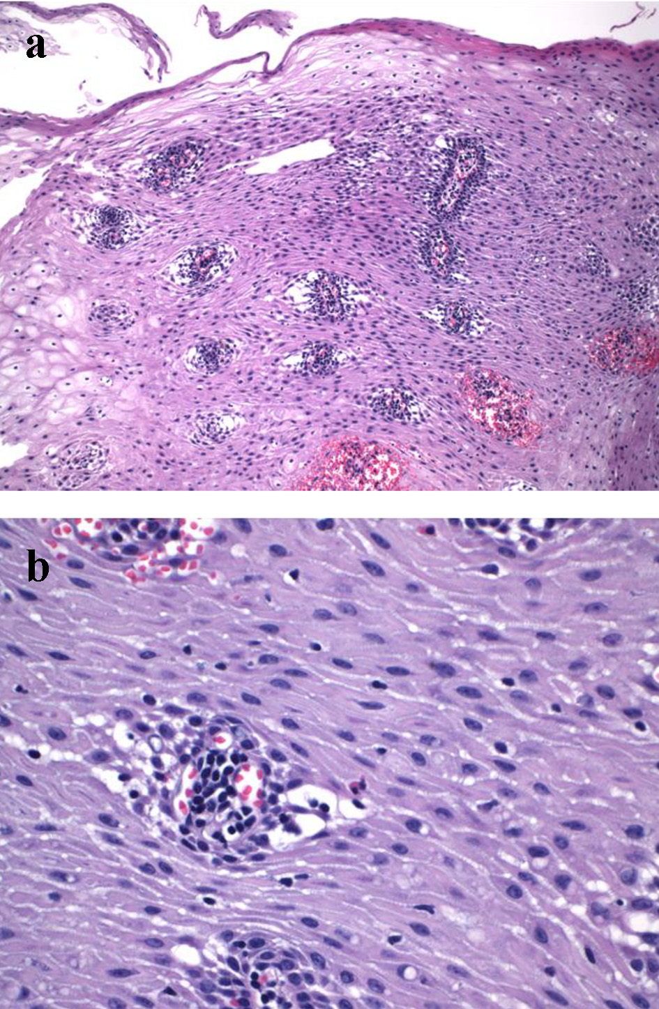
| Peripapillary IEL | IELs in the first five layers of squamous epithelium surrounding the esophageal papillae | Number of IELs per high-power field in the most densely infiltrated fields |
| Interpapillary IEL | IELs in the squamous epithelium beyond the peripapillary regions | Number of IELs per high-power field in the most densely infiltrated fields |
| Neutrophil | Neutrophils in squamous epithelium | Presence or absence |
| Eosinophil | Eosinophils in squamous epithelium | Number of eosinophils per high-power field in the most densely infiltrated fields |
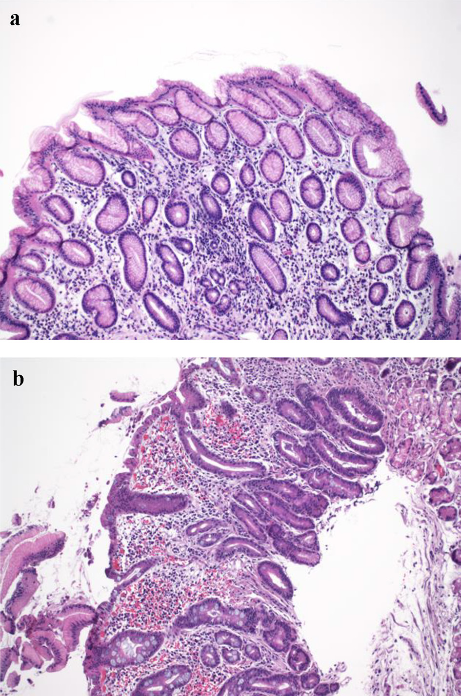
| Columnar | Glandular epithelium | Presence or absence |
| Intestinal metaplasia | Goblet cells | Presence or absence |
| Glandular dysplasia | Neoplastic glandular epithelium confined above the basement membrane | Presence or absence |
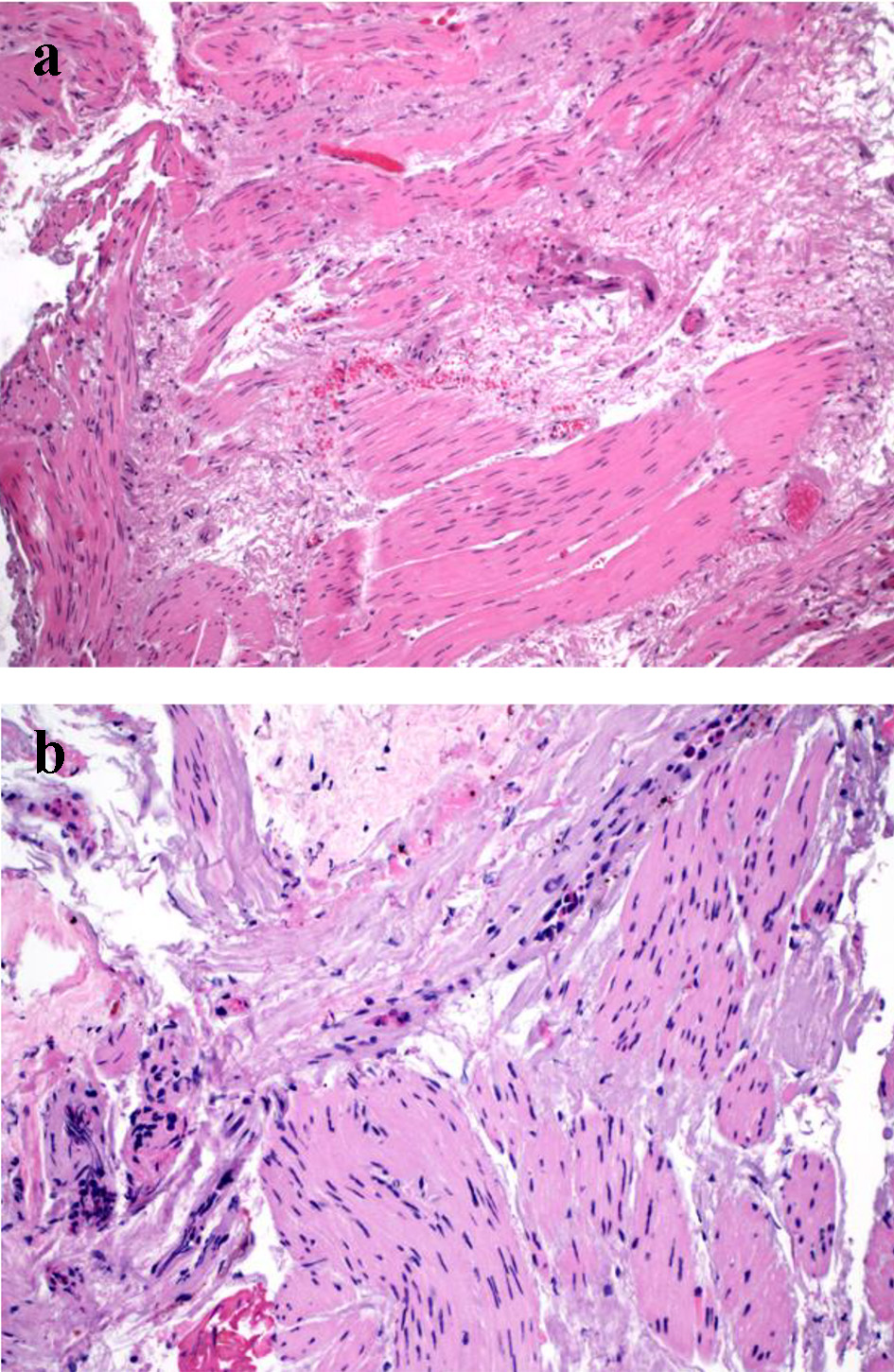
| Subepithelial fibrosis | Increased collagen deposition underneath the epithelium | Presence or absence |
| Submucosal inflammation | Inflammatory cells in the submucosa | Presence or absence |