Gastroenterology Research, ISSN 1918-2805 print, 1918-2813 online, Open Access
Article copyright, the authors; Journal compilation copyright, Gastroenterol Res and Elmer Press Inc
Journal website https://www.gastrores.org

Case Report

Volume 14, Number 2, April 2021, pages 125-128


A Conservative Management of Spontaneously Ruptured Liver Hydatid Cyst

Figures

Figure 1.
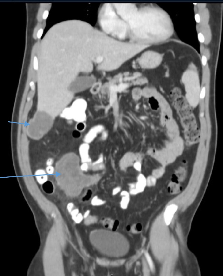
Figure 1. A coronal abdominopelvic CT scan shows hypodense cystic mass in segment 6 of the liver and an additional cystic mass in the mesentery of the small intestine with a moderate amount of free fluid in the abdomen (arrow). CT: computed tomography.
Figure 2.
Figure 2. A coronal abdominopelvic CT scan 4 weeks later demonstrates the same findings of liver hydatid cyst (short arrow) and a well-organized cystic mass at the small-bowel mesentery (long arrow), without free intraabdominal fluid. CT: computed tomography.
Figure 3.
Figure 3. On laparoscopy, a partial pericystectomy of the hydatid cyst in the liver was performed (arrow), following careful cyst aspiration and injection of hypertonic (15%) saline.