Figures
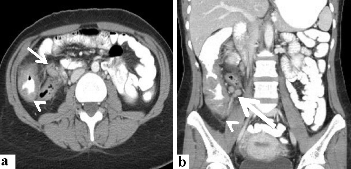
Figure 1. Radiological features of the cecal and lymph nodal basidiobolomycosis (case 1). Abdominal CT scan with intravenous and oral contrast (a) axial, and (b) coronal revealed that diffuse enhancing soft tissue thickening (arrowhead) is seen involving cecum, ascending colon, and an appendix with enlarged enhancing local lymph nodes (arrow). CT: computed tomography.
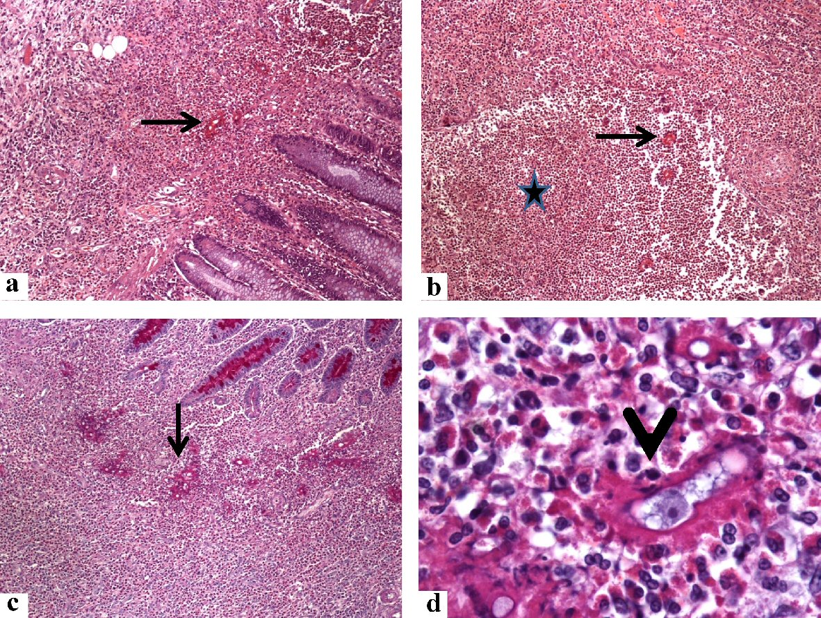
Figure 2. Histological findings in the colonoscopic biopsy specimen from the cecal basidiobolomycosis (case 1). Sections from the cecal ulcerative mass showing some fungal hyphae (arrows) amid dense eosinophil-rich mixed inflammatory cell infiltrate and eosinophilic microabscess (star) in the lamina propria. The B. ranarum appears as hyphae with zygospores. The zygospore resembles trophozoites of amoeba, has foamy cytoplasm with a large nucleolus (arrowhead). The original magnifications are: a, b, c: × 100, and d: × 1,000). B. ranarum: Basidiobolus ranarum.
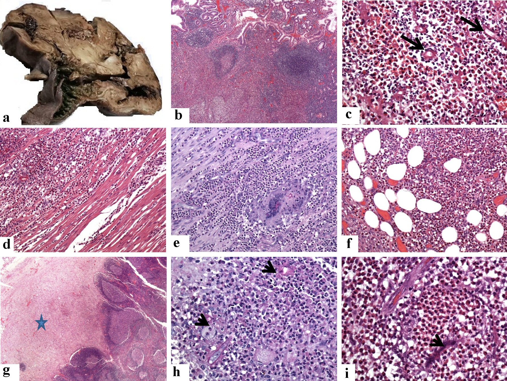
Figure 3. Pathological features of the cecal and lymph nodal basidiobolomycosis (case 1). (a) Grossly, there is diffuse thickening of the wall of the cecum and proximal part of the ascending colon. They are involved by a huge firm mass with a grayish cut section. (b-f) Sections from the cecal mass showing ulceration of the mucosa, increased density of eosinophil-rich mixed inflammatory cell infiltrate in the lamina propria with the formation of multiple lymphoid follicles, around fungal hyphae (arrows, the original magnifications are b: × 40, and c: × 400). This eosinophil-rich mixed inflammation destroys the muscularis propria (d: × 200) with the formation of multinucleated giant cells, phagocytosing the fungal elements (PAS stain, e: × 200), and extending into the subserosal fatty tissue f: × 200). (g-i) Sections show replacement of the subcapsular sinuses (star, g: × 20) of the regional lymph nodes by and multiple fungal structures (PAS stain, arrow, h: × 400). The zygospores resemble trophozoites of amoeba, have foamy cytoplasm with a large nucleolus but no heterochromatin (i: × 1,000, arrowhead), amid eosinophilic microabscess. PAS: Periodic acid-Schiff.
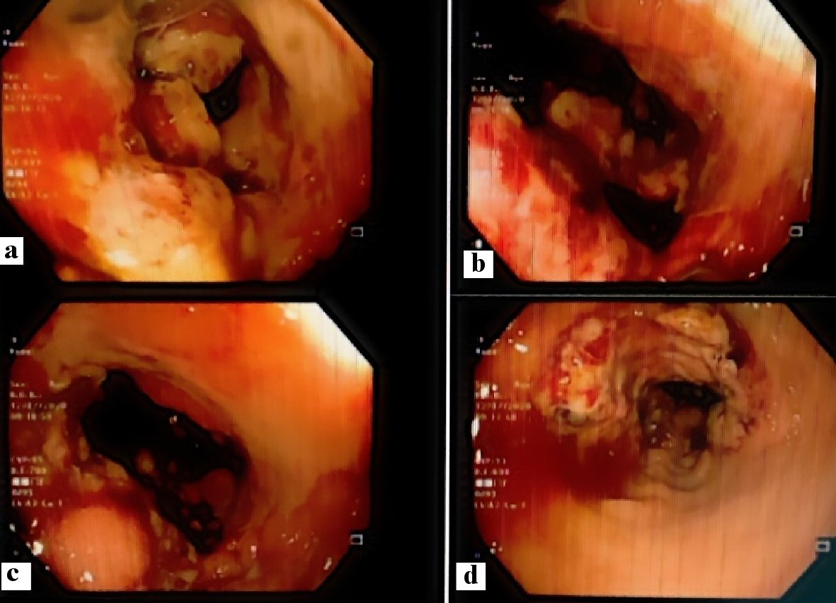
Figure 4. Colonoscopic features of the cecal and appendicular basidiobolomycosis (case 2): (a) Ulcerative mass obstructing the cecum, (b) bleeding ulcer, (c) polypoidal lesion, and (d) narrowing of the colonic lumen.
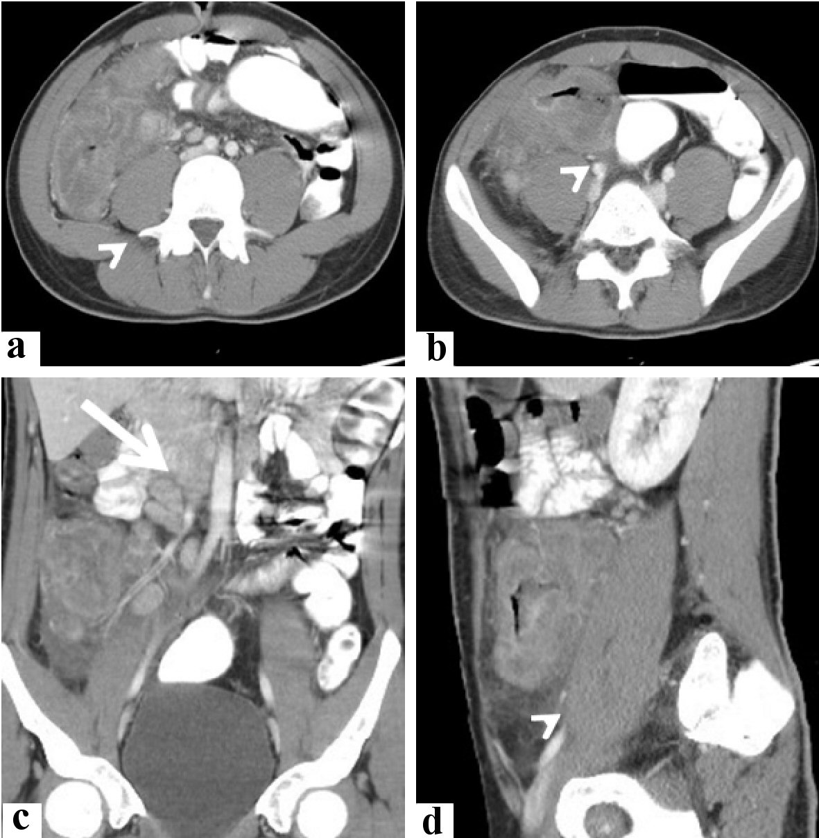
Figure 5. Radiological features of the cecal and appendicular basidiobolomycosis (case 2). Abdominal CT scan with intravenous and oral contrast (a, b) axial, (c) coronal, and (d) sagittal revealed: lobulated enhancing soft tissue mass lesion (arrowhead) is seen involving cecum and ascending colon, contacting abdominal wall and right psoas muscle. It is seen associated with minimal local fluid and enlarged enhancing mesenteric lymph nodes (arrow). CT: computed tomography.
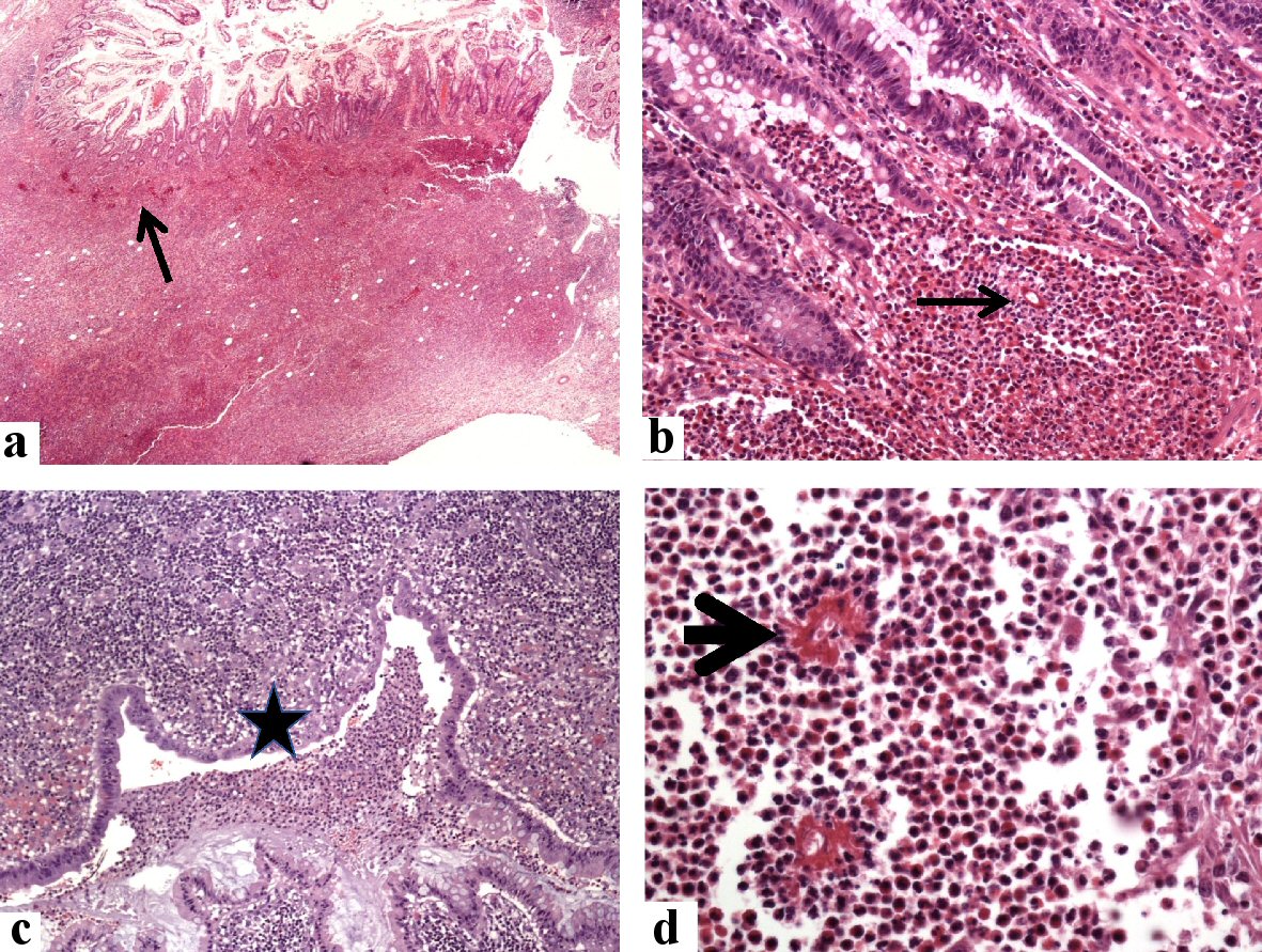
Figure 6. Histological findings in the colonoscopic biopsy specimen from the cecal and appendicular basidiobolomycosis (case 2). Sections from the cecal mass show expansion of the lamina propria by mixed eosinophil-rich inflammatory cells with multiple fungal elements (arrows, hematoxylin and eosin (H&E) stain, a: × 20, and b: × 200), mucosal erosions and luminal necrotic materials with polymorphs (star, c: × 200), and fungal hyphae surrounded by radiating eosinophilic Splendore-Hoeppli bodies (arrowhead, d: × 400).
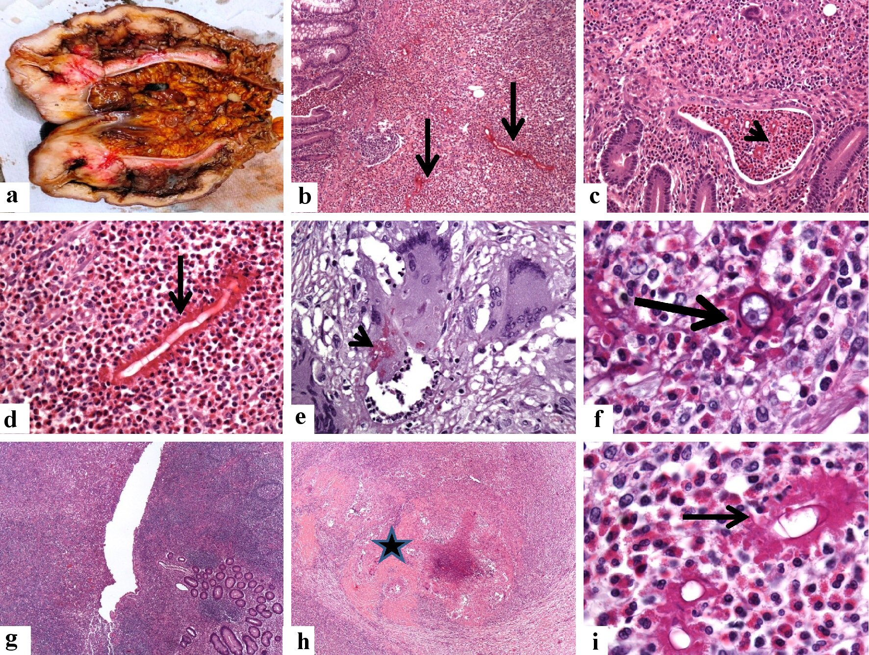
Figure 7. Pathological features of the cecal and appendicular basidiobolomycosis (case 2). (a) En bloc resection of the terminal ileum, appendix, cecum, and ascending colon. There is marked diffuse thickening of the wall of the cecum extending into the proximal part of the ascending colon with obliteration of the lumens. (b-d) The microscopy of the cecal mass reveals a transmural inflammation involving the mucosa (arrow, b: × 100) with the formation of eosinophil crypt abscess (arrowhead, c: × 40), submucosa, of the cecum. The inflammatory reaction consists of eosinophil-rich mixed inflammation contains fungal hyphae surrounded by the eosinophilic Splendore-Hoeppli bodies (d: × 200, black arrow). The Basidiobolus hyphae are thin-walled, septated hyphae, surrounded by eosinophilic material. (e, f) Granulomatous reactions with the formation of giant cells hosting some fungal structures are seen (PAS stain, arrowhead, e: × 400). The zygospores resemble trophozoites of amoeba, have foamy cytoplasm with a large nucleolus but no heterochromatin (f: × 1,000, arrowhead), amid eosinophilic microabscess. (g-i) Sections from the appendix (g: × 40) show transmural necrotizing inflammation (star, h: × 40), composed of eosinophil-rich inflammatory cell infiltrate. The fungal elements are surrounded by the eosinophilic Splendore-Hoeppli bodies (i: × 1,000, thin arrow).