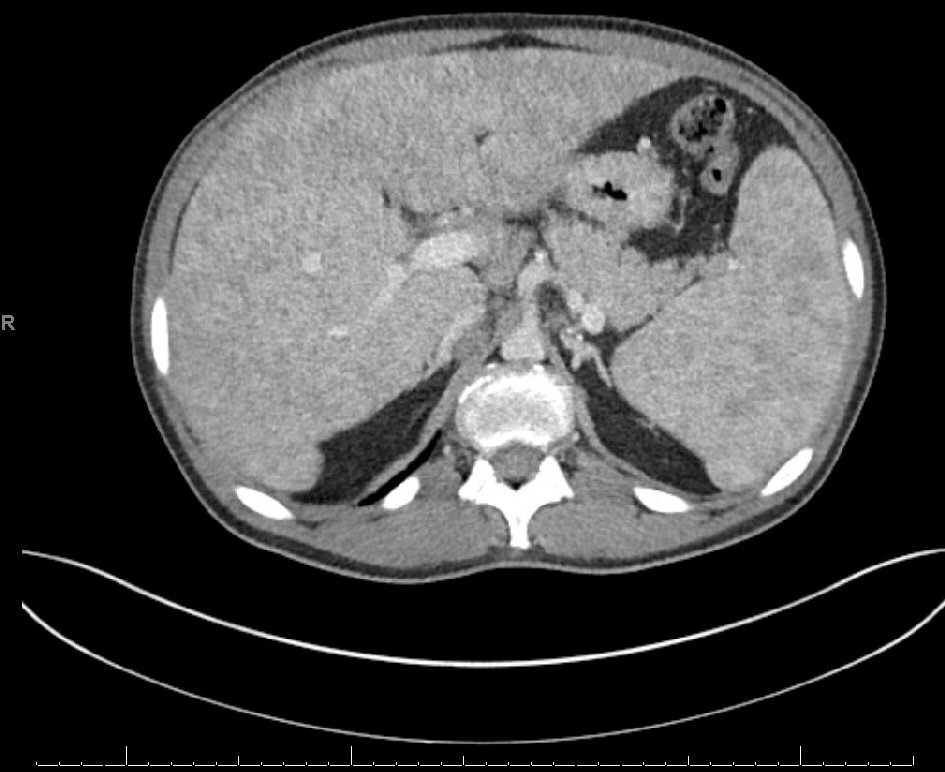
Figure 1. Computed tomography (CT) of the abdomen revealing changes consistent with cirrhosis and splenomegaly.
| Gastroenterology Research, ISSN 1918-2805 print, 1918-2813 online, Open Access |
| Article copyright, the authors; Journal compilation copyright, Gastroenterol Res and Elmer Press Inc |
| Journal website https://www.gastrores.org |
Case Report
Volume 14, Number 2, April 2021, pages 112-115
Sarcoidosis Masquerading as Long-Standing Cholestasis
Figures
Table
| Laboratory investigative marker | Baseline values | Three months after treatment |
|---|---|---|
| Alkaline phosphatase (ALP) | 1,556 U/L | 572 U/L |
| ALP isoenzymes | Liver 76%, bone 24%, intestinal 0%, and placental 0% | N/A |
| Aspartate aminotransferase (AST) | 172 U/L | 41 U/L |
| Alanine aminotransferase (ALT) | 210 U/L | 66 U/L |
| Total bilirubin | 1.9 mg/dL | 1.3 mg/dL |
| Total protein | 7.7 g/dL | 5.9 g/dL |
| Albumin | 3.8 g/dL | 3.1 g/dL |
| Globulin | 3.9 g/dL | 2.8 g/dL |
| International normalized ratio (INR) | 0.92 s | < 0.89 s |
| Alpha-fetoprotein (AFP) | 2.1 ng/mL | N/A |
| Gamma-glutamyl transferase (GGT) | 545 U/L | N/A |
| Carcinoembryonic antigen (CEA) | < 0.5 ng/mL | N/A |
| Liver fibrosis serum panel | Score: 0.73, stage: F3/F4 advanced fibrosis | N/A |
| Vitamin D (1,25(OH)D) | > 156 pg/mL | N/A |
| Model for end-stage liver disease sodium (MELD-Na) | 11 points | 9 points |
| Bronchoalveolar lavage (BAL) cell count | Macrophages 93%, neutrophils 1%, eosinophils 1%, and lymphocytes 6% | N/A |