Gastroenterology Research, ISSN 1918-2805 print, 1918-2813 online, Open Access
Article copyright, the authors; Journal compilation copyright, Gastroenterol Res and Elmer Press Inc
Journal website https://www.gastrores.org

Original Article

Volume 14, Number 1, February 2021, pages 13-20


Characterization of Chronic Gastritis in Lynch Syndrome Patients With Gastric Adenocarcinoma

Figure

Figure 1.
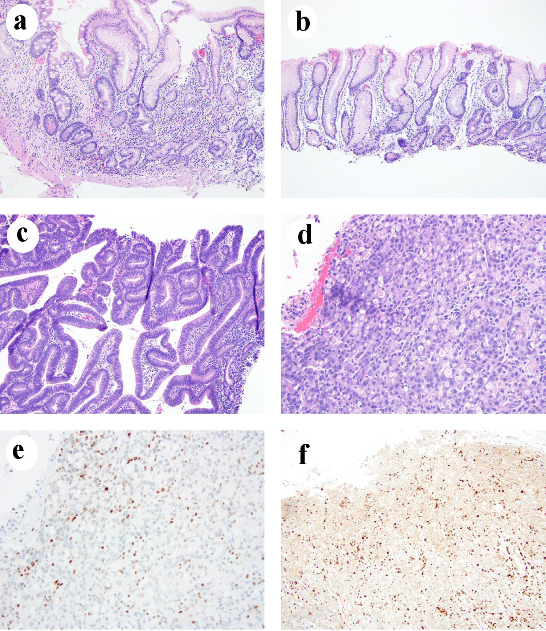
Figure 1. Chronic gastritis in one LS patient who developed gastric adenocarcinoma. (a) Body mucosa showing chronic inactive atrophic gastritis with intestinal metaplasia and pseudopyloric gland metaplasia (H&E stain, 100 ×). (b) Antral mucosa showing mild chronic inactive gastritis without intestinal metaplasia (H&E stain, 100 ×). (c) Histomorphology of low-grade dysplasia (H&E stain, 100 ×). (d) Signet ring cell/diffuse type carcinoma (H&E stain, 200 ×). (e) Loss of MLH1 in gastric adenocarcinoma nuclei (immunohistochemical stain, 200 ×). (f) Loss of PMS2 expression in gastric adenocarcinoma nuclei (immunohistochemical stain, 100 ×). H&E: hematoxylin and eosin; LS: Lynch syndrome.

Tables

Table 1. Clinicodemographics and Gastric Tumor Characteristics of LS Patients in This Study
 
Number of cases with information availableResults
Lynch syndrome.
Age, mean (range), years1163 (23 - 83)
Personal cancer history1110 (90.9%) had personal cancer history including:
Colon (6)/pancreas (1)/duodenum (1)
Breast (1)/endometrium (1)
Non-melanoma skin cancer (1)
Medulloblastoma (1)
Malignancy, not other specified (1)
Family history of colon cancer76 (85.7%)
Family history of gastric cancer90 (0%)
Symptoms11Symptomatic: 5
  Abdominal pain (2)/epigastric discomfort (1)
  Reflux (1)/anemia (1)
Symptomatic and in surveillance program: dysphagia (1)
Asymptomatic and in surveillance program: 5
Tumor location12Fundus: 3
Body: 5
Fundus and body: 1
Antrum: 2
Tumor histology type11Intestinal or mixed: 7 (63.6%)
Signet ring cell/diffuse: 4 (36.4%)
Depth of invasion11Intramucosal: 4
Submucosal: 3
Muscularis propria: 2
Subserosal or deeper: 2
Nodal metastases80 (0%)
Distant metastases110 (0%)
Loss of mismatch repair proteins6MLH1 loss: 3
MSH2 loss: 2
Unknown: 1
Precursor lesion11Low-grade dysplasia: 3
High-grade dysplasia: 3


Table 2. Background Gastric Mucosal Changes in LS Patients With Gastric Adenocarcinoma
 
Number of cases with information availableResults
H. pylori: Helicobacter pylori; LS: Lynch syndrome; IM: intestinal metaplasia; ECL: enterochromaffin-like.
Body or antral biopsy11Chronic gastritis: 8 (72.7%)
  Chronic H. pylori-negative gastritis: 7
  Chronic H. pylori gastritis: 1
Atrophy: 5 (45.4%)
IM: 4 (36.3%)
Body biopsy8Moderate chronic gastritis: 5 (63%)
  Chronic H. pylori-negative gastritis: 4
  Chronic H. pylori gastritis: 1
Atrophy: 4 (50%)
Pseudopyloric gland metaplasia: 4 (50%)
IM: 3 (38%)
ECL cell hyperplasia: 3 (37.5%)
Normal histology: 3 (37%)
Antral biopsy8Moderate chronic gastritis: 2 (25%)
Mild chronic gastritis: 2 (25%)
  Mild chronic H. pylori-negative gastritis with IM: 1
  Mild chronic H. pylori gastritis: 1
IM only: 1 (12.5%)
Reactive gastropathy: 1 (12.5%).
Normal histology: 2 (25%)
Body and antral biopsy5Body predominant chronic gastritis with IM: 2 (60%)
Body predominant chronic gastritis without IM: 1 (20%)
Body predominant H. pylori gastritis with IM: 1 (20%)
Normal histology: 1 (20%)


Table 3. Comparison of Gastric Mucosal Changes Between LS Patients With Gastric Cancer (Current Study) and Without Gastric Cancer From One Previous Study
 
Chronic gastritis, n (%)H. pylori infection, n (%)Atrophy, n (%)IM, n (%)
*Data were from previously published study [8]. H. pylori: Helicobacter pylori; LS: Lynch syndrome; IM: intestinal metaplasia.
LS patients with gastric adenocarcinoma8 (75)1 (9.1)5 (45.4)4 (36.3)
LS patients without gastric adenocarcinoma (n = 73)*23 (31.5)19 (26.0)10 (13.7)10 (13.7)
P value0.0160.440.0220.08