Gastroenterology Research, ISSN 1918-2805 print, 1918-2813 online, Open Access
Article copyright, the authors; Journal compilation copyright, Gastroenterol Res and Elmer Press Inc
Journal website https://www.gastrores.org

Case Report

Volume 14, Number 1, February 2021, pages 41-44


Ampullitis Superimposed Obstructive Jaundice in a Patient With COVID-19

Figures

Figure 1.
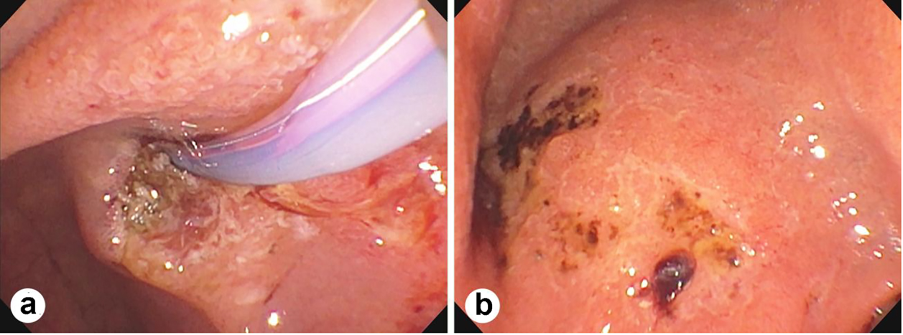
Figure 1. Endoscopic retrograde cholangiopancreatography (ERCP) showing duodenal major papillae and ampullary ulceration (a) and multiple ulcers in the duodenal bulb with bleeding (b).
Figure 2.
Figure 2. Duodenal major papillae and ampullary biopsy showing mucosal ulceration (a, b, arrows), vasculitis (c, left, arrows), thrombi (c, right arrows), smudge nuclei (c, left, and d, arrow heads) and ganglions involved by the inflammation (d, arrows) (a-d, H&E stains; a, × 100; b-d, × 400).
Figure 3.
Figure 3. Bile duct brushing specimen showing clusters of atypical epithelial cells with high-grade dysplasia (Papanicolaou stain, × 400).
Figure 4.
Figure 4. Chest X-ray showing mild hazy density within the lower lung lobes bilaterally without focal consolidation.