Gastroenterology Research, ISSN 1918-2805 print, 1918-2813 online, Open Access
Article copyright, the authors; Journal compilation copyright, Gastroenterol Res and Elmer Press Inc
Journal website https://www.gastrores.org

Case Report

Volume 13, Number 6, December 2020, pages 279-282


A Case of A Mesenteric Cyst Mimicking a Biloma

Figures

Figure 1.
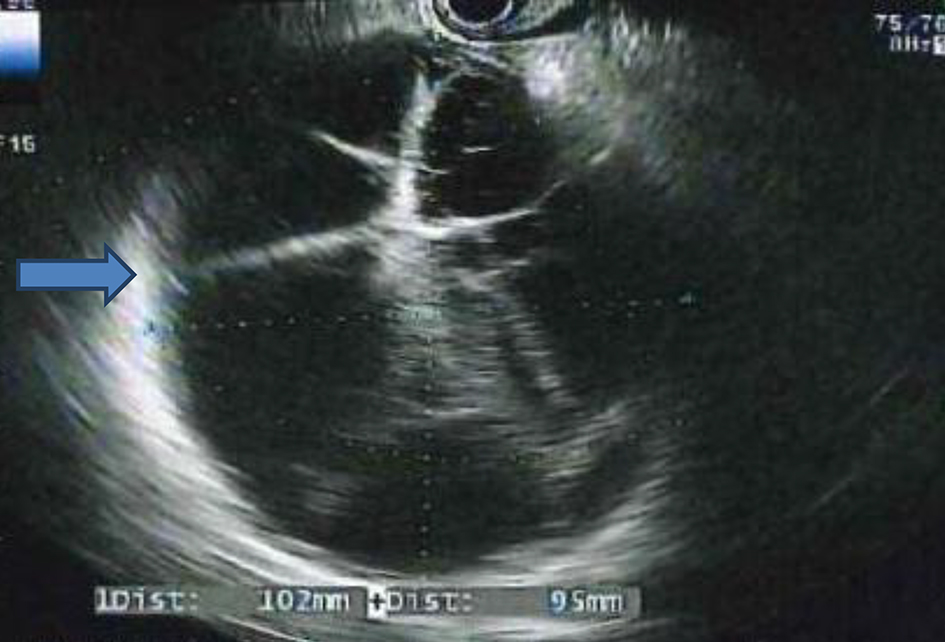
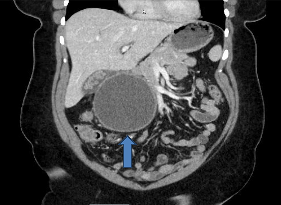
Figure 1. Abdominal computed tomography (CT) scan showing a 10.3 × 9.8 × 11.3 cm right upper quadrant (RUQ) cystic mass (blue arrow).
Figure 2.
Figure 2. Endoscopic ultrasonography (EUS) revealing a large septated mass (blue arrow).
Figure 3.
Figure 3. Intraoperative cholangiogram showing lack of dye extravasation between the mass and biliary tree.
Figure 4.
Figure 4. Prominent lymphoid aggregate surrounding small vessels and capillaries within the cyst wall (mid-power views, × 20).
Figure 5.
Figure 5. Foamy histocytes surrounding blood vessels within the cyst wall (high power, × 40).
Figure 6.
Figure 6. Pigmented and foamy macrophages near the cyst lining (high power, × 40).