Gastroenterology Research, ISSN 1918-2805 print, 1918-2813 online, Open Access
Article copyright, the authors; Journal compilation copyright, Gastroenterol Res and Elmer Press Inc
Journal website https://www.gastrores.org

Original Article

Volume 13, Number 4, August 2020, pages 129-137


Primary Localized Amyloidosis of the Intestine: A Pathologist Viewpoint

Figures

Figure 1.
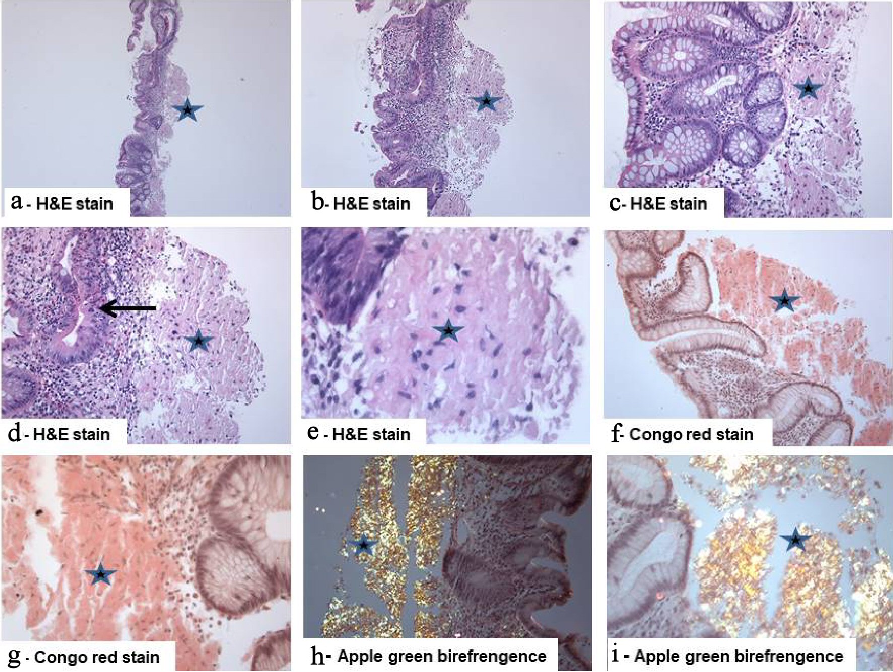
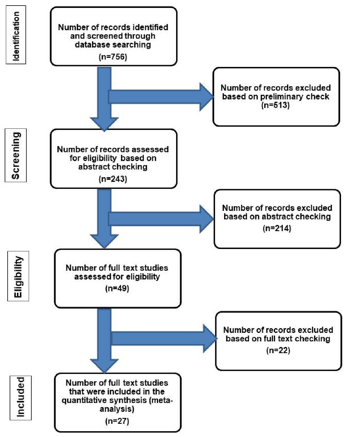
Figure 1. Flow chart of literature search and study selection for cases of localized intestinal amyloidosis. The inclusion criteria included patients who had amyloid deposition in the intestine without evidence of systemic involvement or an associated condition leading to secondary amyloidosis [42]. The exclusion criteria included patients with one or more of the followings: 1) Evidence of chronic diseases such as collagen vascular disease, that may have led to secondary amyloidosis [42]; 2) Additional organ involvement by amyloidosis; 3) Intestinal symptoms suggestive of amyloidosis but without confirmatory diagnostic tissue biopsy; 4) Plasma cell dyscrasia, myeloma, lymphoplasmacytic disorders or other B-cell malignancies such as Waldenstrom’s macroglobulinemia [5]; 5) Systemic AL type [43]; 6) Laboratory findings revealing positive detection of monoclonal light chains in serum and/or urine [15].
Figure 2.
Figure 2. Isolated primary amyloidosis of the intestine (case report): (a - e) Colonic mucosa with deposition of pale eosinophilic amorphous material (star) in the submucosa with abundant deposits of pink amorphous materials with some cracking artifacts, characteristic of amyloid. Focal surface mucosal ulceration is noted. The background mucosa shows focal active inflammation, including cryptitis (arrow), and there is patchy crypt distortion. No granulomas are seen (hematoxylin and eosin: (a) × 20, (b) × 40, (c, d) × 200, and (e) × 400). (f, g) Congo red stain which reveals the characteristic salmon-pink color, confirming the presence of amyloid deposits (Congo red, (f) × 200 and (g) × 400). (h, i) The deposits exhibit characteristic apple-green birefringence under polarized light (Congo red under polarized light ((h) × 200 and (i) × 400).

Tables

Table 1. Data From Individual Studies of Primary Localized Amyloidosis of the Small Intestine
 
CaseSexAge (years)SitesClinical manifestationsColonoscopic findingsStudies
1F44IleumAcute abdominal pain, diarrhea, and vomitingThickening of the wall of the ileum and ascending colon[28]
2M47JejunumPseudo-intestinal obstructionThickening of the mucosal folds and multiple polypoid protrusions[29]
3F81JejunumMelena, and anemiaDiffuse thickening of the mucosa with friability, nodularity, erosions, and polypoid protrusions[30]
4M71Small intestineIntestinal obstructionDiffuse thickening of the wall of the small intestine[31]
5F51Jejunum and ileumChronic diarrhea and malabsorptionThickening of the jejunal and proximal ileal mucosal folds[32]
6MMiddle ageSmall intestineAcute abdomenPerforation of the wall[33]
7M62Duodenum and jejunumAbdominal pain, nausea, and constipationMultiple shallow ulcers and several erosions[34]
8M62Small intestineSmall bowel obstructionBand of amyloid and connective tissue surrounding the small intestine[35]
9MAgedSmall intestineAbdominal pains and hematemesisMucosal ulcerations with discrete nodules[36]
10M60Small intestineNonspecific digestive symptomsMultiple mucosal polyps[37]
11M74JejunumNonspecific digestive symptomsPolypoid, pseudo-tumoral mucosal formations.[38]
12F68Small intestineNonspecific digestive symptomsThickening of the wall with rough, and polypoid intestinal mucosal folds[39]
13F, M59Small intestine (three cases)Heartburn and constipation-[6]
14M55DuodenumScreening for cancer stomachA localized depressed lesion[40]


Table 2. Data From Individual Studies of Primary Localized Amyloidosis of the Colon
 
CaseSexAge (years)SitesClinical presentationColonoscopic findingsStudies
1M52Transverse colonA periumbilical pain, rectal bleeding, and weight lossUlcerations and luminal narrowing by multiple polypoid lesions[15]
2MElderlyTransverse colonIntestinal obstructionA stenosing mass[16]
3MMiddle ageTransverse colonNonspecific digestive symptomsSubmucosal mass[17]
4M74Descending colonAnemiaUlcerative lesions[18]
5M88Sigmoid colonAcute abdominal pain, rectal bleeding, and fecal peritonitisMucosal ulceration, perforation, and thickening of the bowel wall[20]
6F64Sigmoid colonHematocheziaA submucosal tumor-like mass[21]
7M51Sigmoid colonHeme-positive stoolsMucosal ulcer[22]
8M58Sigmoid colonRectal bleedingA single friable, rounded mucosal lesion[23]
9M73Sigmoid colonRoutine colonoscopyA 1.5-cm shallow depressed mucosal lesion[24]
10F79SigmoidNonspecific digestive symptomsSeveral irregularly-shaped discrete ulcerations[25]
11M46Descending colonAbdominal pain, bloating, flatulence and hematocheziaHemorrhagic mucosa and amass lesion[12]
12F59Ascending and descending colonsRectal bleedingDiffuse nodular friable lesions (ascending colon) and several irregular large ulcers with nodularity (descending colon)[26]
13F, MMiddle ageColonRectal bleeding (two cases)Thickening of the wall and mucosal ulcerations[27]
14F, M59Colon (seven cases)Heartburn and constipation-[6]
15F44Ascending colonAcute abdominal pain, diarrhea, and vomitingThickening of the wall of the ileum and ascending colon[28]


Table 3. Clinical, Colonoscopic and Histological Findings in the Primary Localized Amyloidosis of the Intestine
 
AspectsSmall intestine (duodenum, jejunum, and ilium)Colorectum
Age (mean ± SEM)62 ± 3.7 years [6, 28-39]65.1 ± 3.9 years [6, 12, 15-27]
Male to female ratio8/412/3
Site of involvement
Jejunum (40%)The left colon (44%)
Ileum (7%)Transverse colon (11%)
Clinical presentations
Abdominal pain and intestinal obstruction (33%) [6, 28, 29, 31, 33-36]Rectal bleeding (37%) [15, 20-23], followed by abdominal pain (11%) [15, 20] and anemia [18]
Colonoscopic findings
Thickening of the wall (18%) [28-32, 39]; polyps and polypoid mucosal protrusions (11%) [37-39]; ulcerations (7%) [34, 36]; friability and nodularity of the mucosa (7%) [30, 36], and perforation (3%) [33].Mass lesions (tumor-like lesion, polypoid protrusions, and polyps) with narrowing were the most common (29%) [12, 15-18, 21]. Ulcerations with solitary or multiple ulcers (14%) [20, 22, 25-27], thickening of the wall (7%) [20, 27], nodularity and friability of the mucosa (2%) [23, 24, 26], and perforation (3%) [20].
Histological features
Amyloid deposits; increased density of mixed inflammatory cells in the lamina propria; light chain protein [21, 34].Amyloid deposits; increased density of mixed inflammatory cells in the lamina propria; foreign body giant cell reaction; amyloid angiopathy; necrotizing angiitis; focal active colitis; and immunoglobulin light-chain (AL) [16, 23, 24].