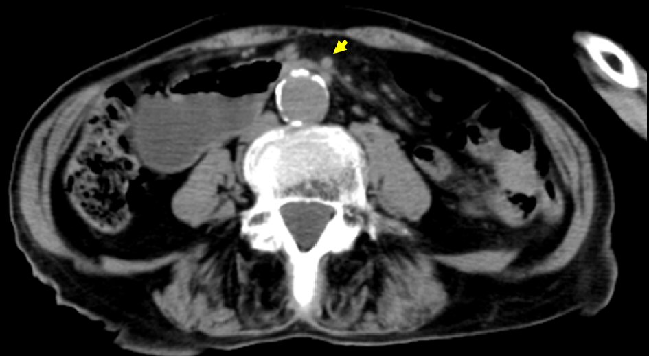
Figure 1. Abdominal CT revealed dilatation of the duodenum up to the level of the distal third portion, which was sandwiched between the aorta and SMA (arrow). CT: computed tomography.
| Gastroenterology Research, ISSN 1918-2805 print, 1918-2813 online, Open Access |
| Article copyright, the authors; Journal compilation copyright, Gastroenterol Res and Elmer Press Inc |
| Journal website http://www.gastrores.org |
Case Report
Volume 12, Number 6, December 2019, pages 320-323
Endoscopic Gastrojejunostomy for Superior Mesenteric Artery Syndrome Using Magnetic Compression Anastomosis
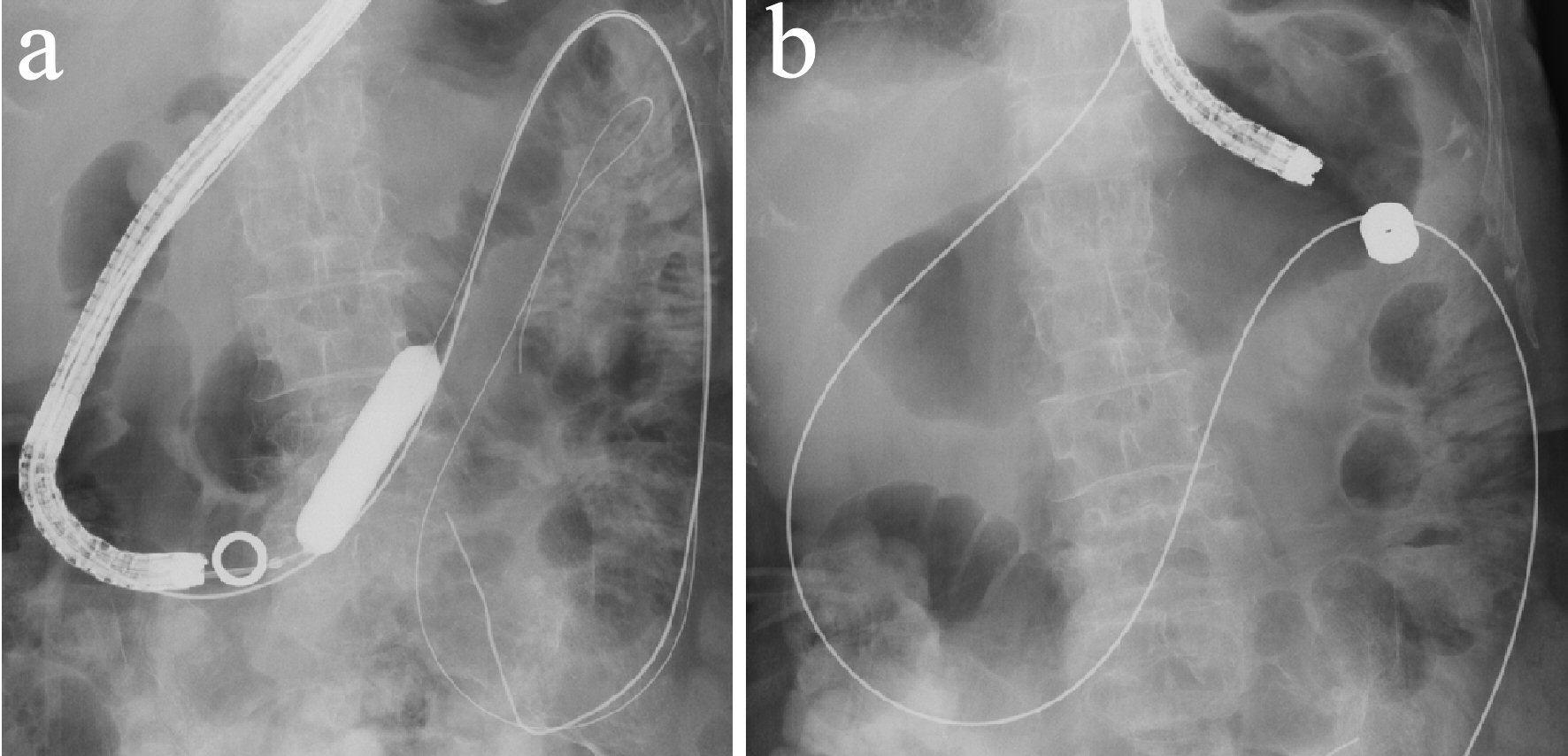
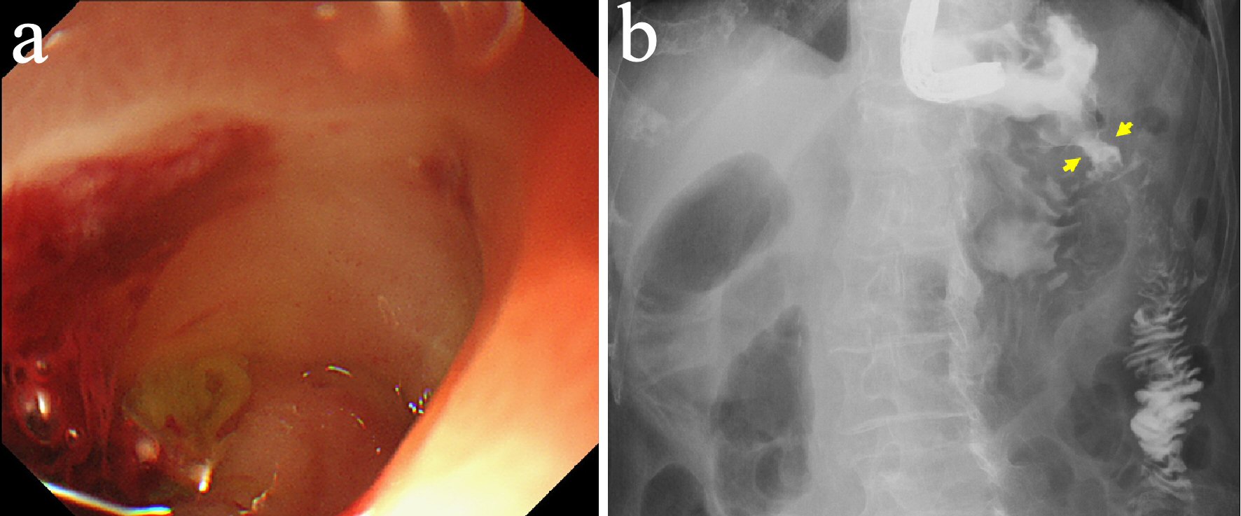
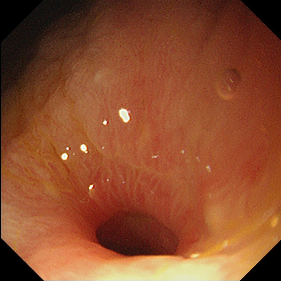
Figures