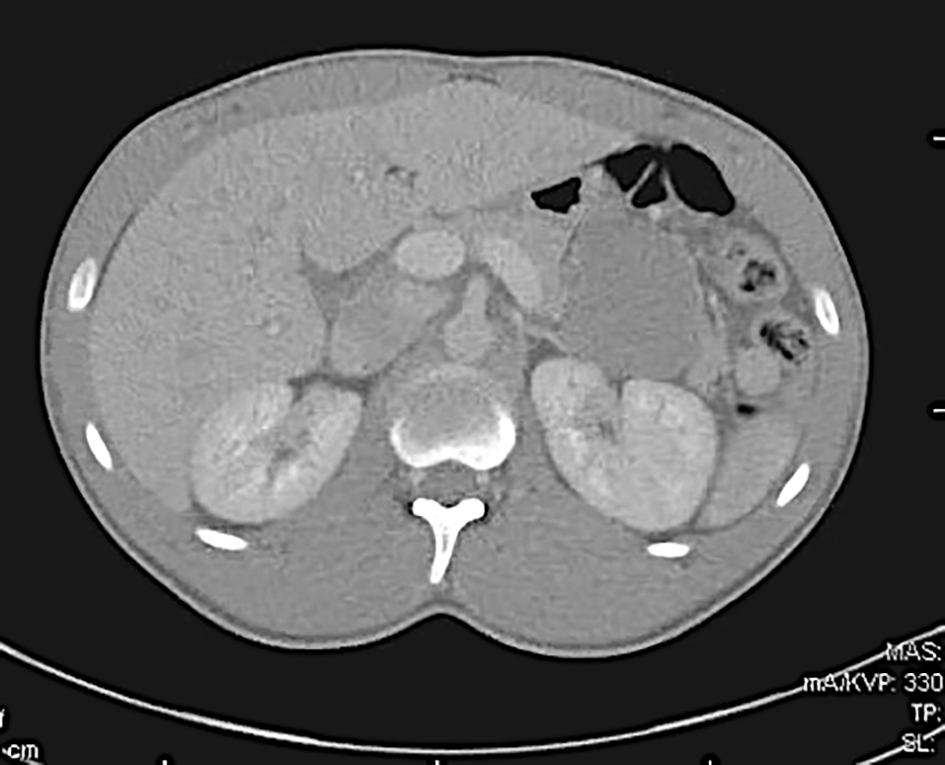
Figure 1. Contrast-enhanced computed tomography evidencing 5.8 cm pancreatic mass.
| Gastroenterology Research, ISSN 1918-2805 print, 1918-2813 online, Open Access |
| Article copyright, the authors; Journal compilation copyright, Gastroenterol Res and Elmer Press Inc |
| Journal website http://www.gastrores.org |
Case Report
Volume 12, Number 3, June 2019, pages 174-175
Tackling the Diagnosis: Solid Pseudopapillary Tumor of the Pancreas in a Young Man
Figures