| Clinical setting | Mass, ill-defined lesion or abnormal imaging finding | Random biopsy from patients with or without metabolic risk factors, with or without liver function abnormalities | Mass or ill-defined lesion | Mass, ill-defined lesion or abnormal imaging finding, with or without a central scar |
| Macroscopic findings | Mass, ill-defined lesion or vague abnormalities with gold-yellow appearance | Non-specific | Mass or ill-defined lesion with cut surface different from adjacent liver | Mass or ill-defined lesion with or without a central scar |
| Histology | | | | |
| Relationship to adjacent liver parenchyma | Variable (well demarcated to focally invasive into adjacent portal tracts) | Non-invasive | Variable (well demarcated or invasive into adjacent portal tracts) | Non-invasive |
| Central scar | Variable | Absent | Absent | Present in large lesions but variable in small lesions |
| Portal tracts | Absent | Present | Absent | Absent |
| Ductular reaction | Absent | Variable but present in most cases | Absent | Present in fibrous septa |
| Thick-walled vessels within fibrous bands | Absent | Absent | Absent | Present |
| Fibrous bands | Variable | Variable | Variable | Present |
| Isolated arteries immediately in contact with hepatocytes | Present | May be present (small) | Present | Absent |
| Small cell change | Variable | Absent | Present in many cases | Absent |
| Increased nuclear/cytoplasmic ratio | Variable | Absent | Present | Absent |
| Pleomorphism | Variable (minimal to marked) | Minimal to mild | Variable (mild to marked) | Minimal to mild |
| Acinar/rosette formation | Absent to minimal | Absent | Variable (absent to marked) | Variable (small rosettes, focal) |
| Mitotic figures | Variable (can be rare) | Absence | Variable (usually identifiable) | Absence |
| Capillarization of endothelium | Variable | Absence | Presence | Absence |
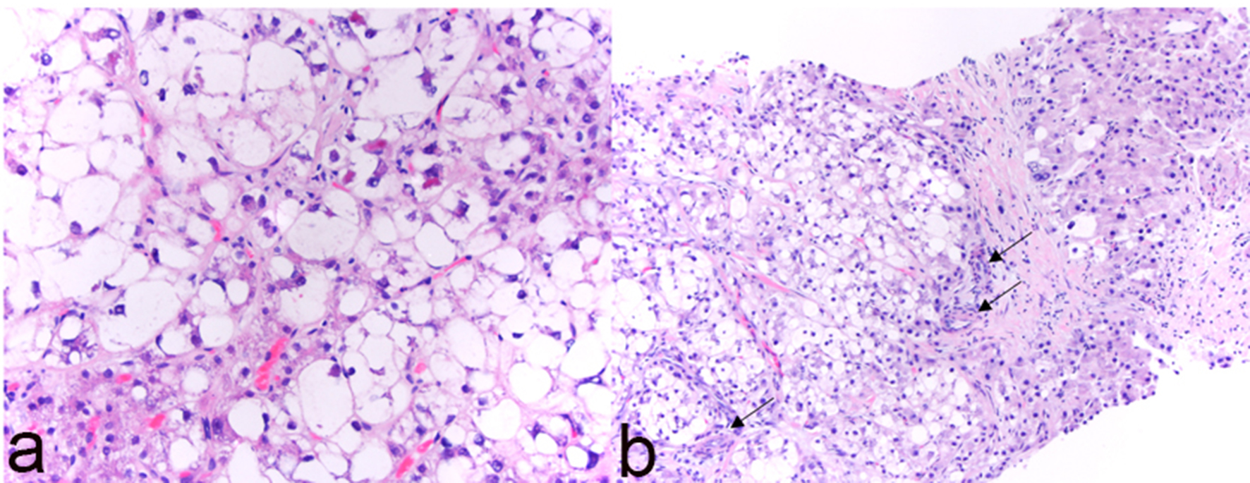
| Steatosis | Presence | Presence | Absence | Presence |
| Hepatocyte ballooning | Presence | Presence | Absence | Presence |
| Mallory-Denk Bodies | Presence | Presence | Absence | Presence |
| Inflammation | Presence (mild to severe) | Presence (mild to severe) | Variable | Variable |
| Thick plates > 3 hepatocytes | Variable presence | Absence | Variable presence | Variable presence (limited in extent, focal) |
| Reticulin fiber | May be lost | May be slightly attenuated | Lost in most cases | May be slightly attenuated |
| Immunostain | | | | |
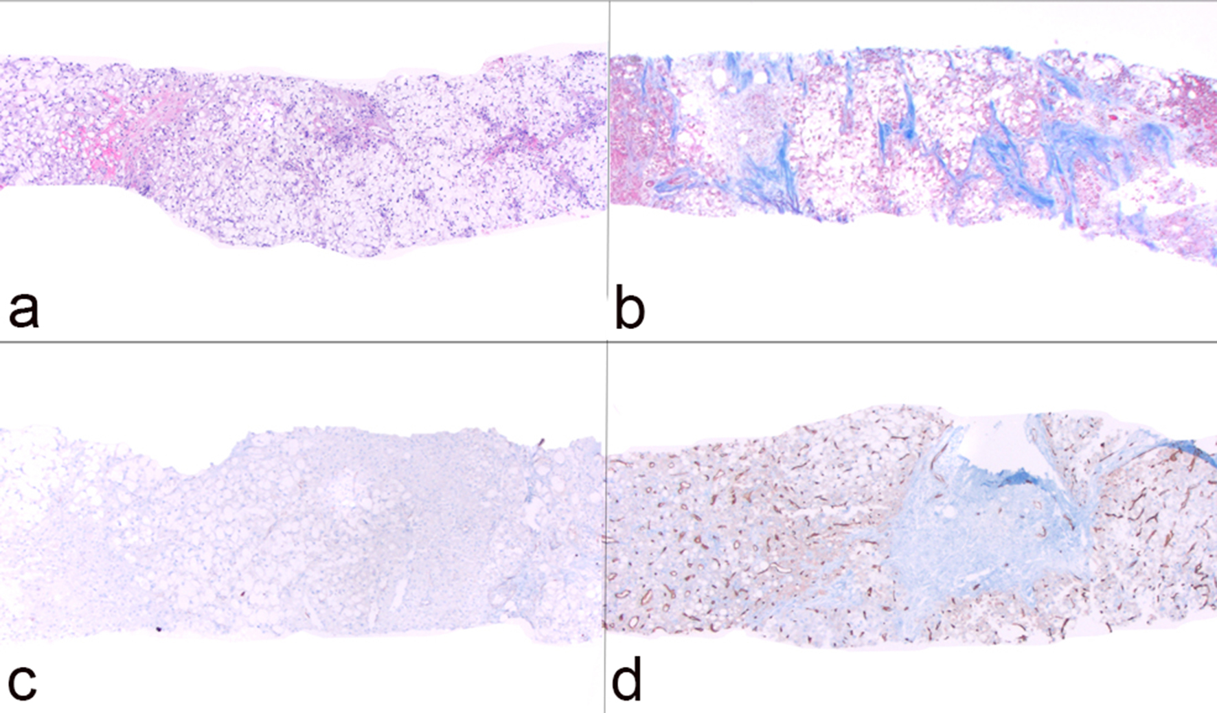
| Diffuse sinusoidal CD34 staining | Presence in most cases | Absence | Presence | Absence |
| Glypican-3 | Variable presence (cytoplasmic and canalicular) | Absence | Presence in many cases | Absence |
| Diffuse glutamine synthetase | Variable presence (cytoplasmic staining with perinuclear accentuation) | Absence | Presence in many case | Absence* |
| Nuclear β-catenin | Presence in 6% cases | Absence | Presence in some cases | Absence |