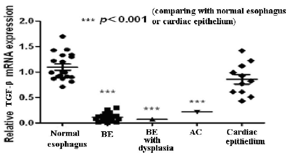
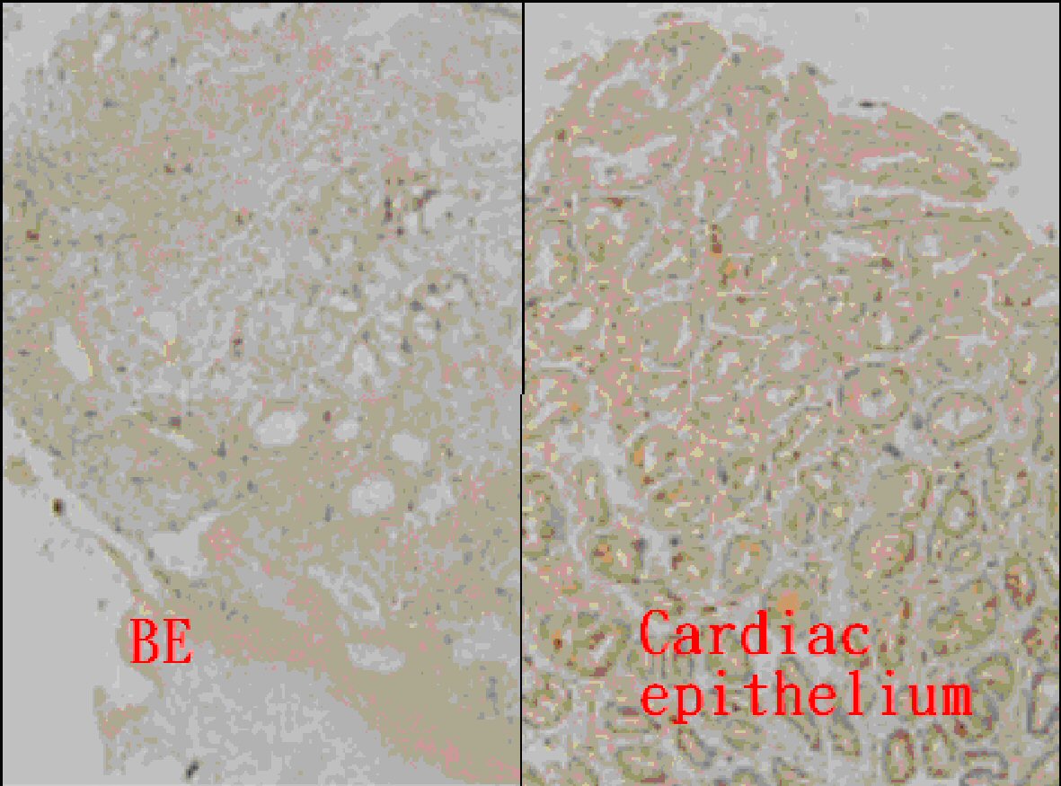
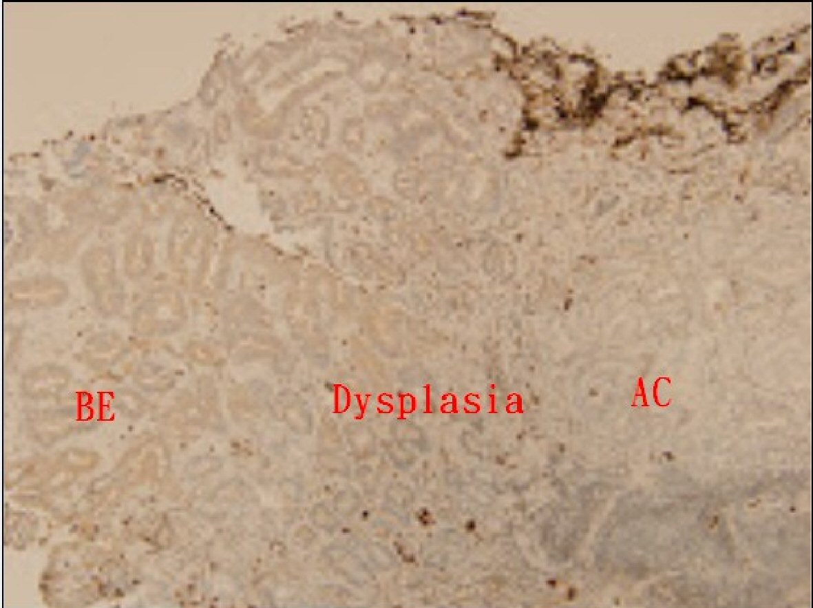
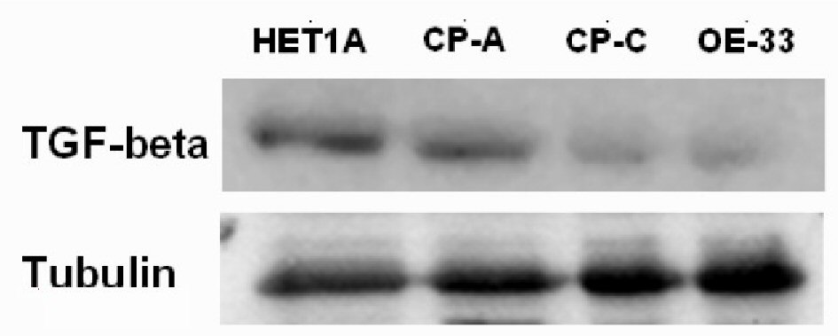
Figure 1. The value of TGF-β mRNA in the normal esophagus, Barrett’s esophagus (BE), BE with dysplasia, adenocarcinoma (AC), and gastric cardiac epithelium.
| Gastroenterology Research, ISSN 1918-2805 print, 1918-2813 online, Open Access |
| Article copyright, the authors; Journal compilation copyright, Gastroenterol Res and Elmer Press Inc |
| Journal website http://www.gastrores.org |
Original Article
Volume 11, Number 3, June 2018, pages 189-194
Low Expression of Transforming Growth Factor β in the Epithelium of Barrett’s Esophagus
Figures
Tables
| Basic characteristic | |
|---|---|
| AC: adenocarcinoma; BE: Barrett’s esophagus; BMI: body mass index; LSBE: long-sequment Barrett’s esophagus; SSBE: short-sequment Barrett’s esophagus. | |
| Age (year) | 62.00 ± 16.64 |
| Gender (male) | 23 (76.7%) |
| Body weight (kg) | 66.78 ± 11.47 |
| BWI (kg/m2) | 24.69 ± 3.28 |
| Waist (cm) | 90.27 ± 9.40 |
| Endoscopic findings | |
| SSBE/LSBE | 26 (86.7%)/4 (13.3%) |
| Erosive esophagitis | 12 (40.0%) |
| L.A. grade A/B/C/D | 6/4/2/0 |
| Hiatal hernia | 12 (40.0%) |
| H. pylori infection | 2 (6.7%) |
| Histological findings | |
| BE | 28 (93.4%) |
| BE with dysplasia | 1 (3.3%) |
| AC | 1 (3.3%) |
| IHC stain | 0 | 1+ | 2+ | 3+ | Total |
|---|---|---|---|---|---|
| GEJ: gastroesophageal junction; IHC: immunohistochemical. | |||||
| GEJ | 10 | 8 | 4 | 1 | 23 |
| Cardiac epithelium | 1 | 0 | 4 | 18 | 23 |