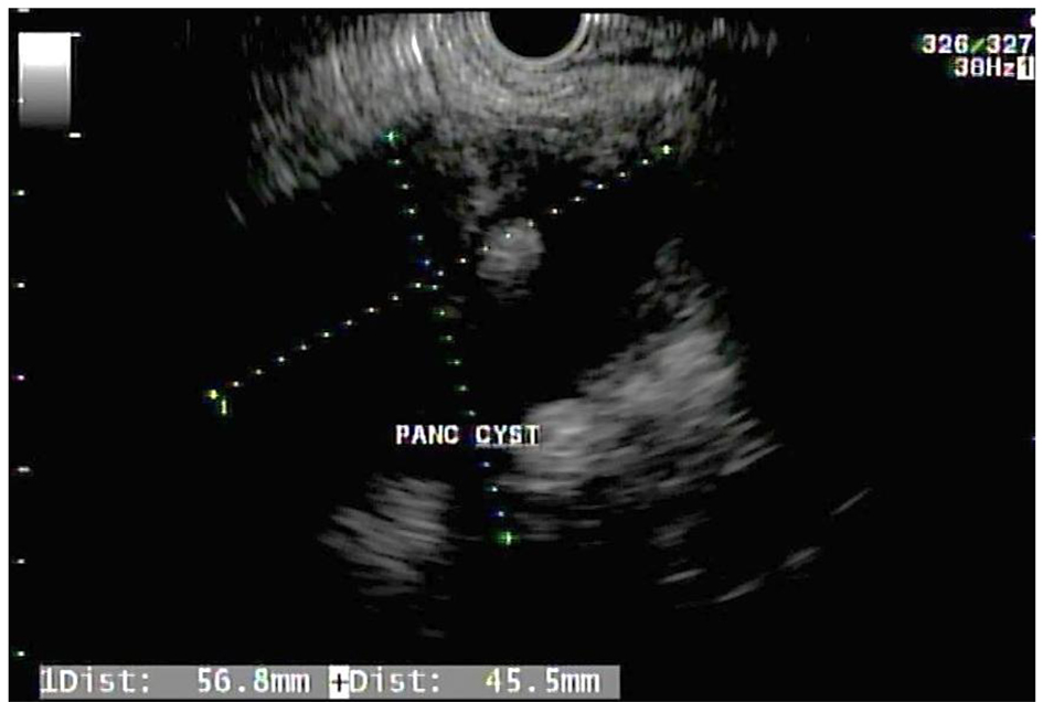
Figure 1. Endoscopic ultrasound showing a large heterogeneous cystic lesion with hypoechoic wall layers measuring 56.8 × 45.5 mm in the tail of the pancreas.
| Gastroenterology Research, ISSN 1918-2805 print, 1918-2813 online, Open Access |
| Article copyright, the authors; Journal compilation copyright, Gastroenterol Res and Elmer Press Inc |
| Journal website http://www.gastrores.org |
Case Report
Volume 10, Number 5, October 2017, pages 322-324
Large Dermoid Cyst Presenting as Recurrent Pancreatitis
Figures