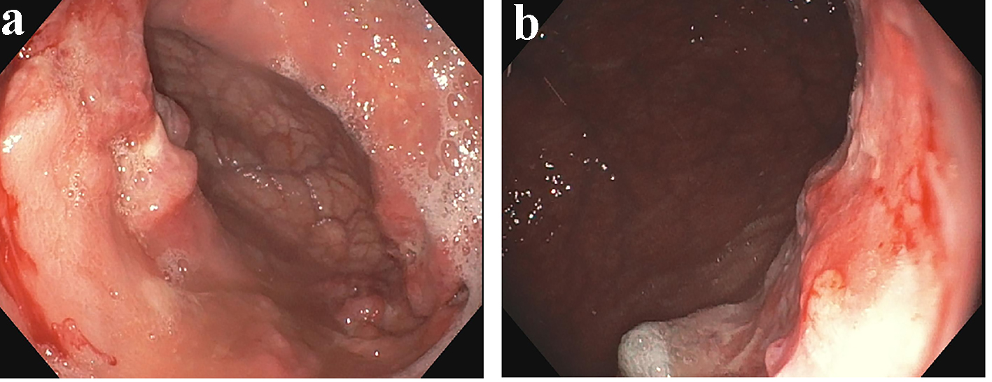
Figure 1. (a, b) A diffuse gastric body mass.
| Gastroenterology Research, ISSN 1918-2805 print, 1918-2813 online, Open Access |
| Article copyright, the authors; Journal compilation copyright, Gastroenterol Res and Elmer Press Inc |
| Journal website http://www.gastrores.org |
Case Report
Volume 10, Number 5, October 2017, pages 311-314
Concurrent Primary Gastric and Pulmonary Diffuse Large B-Cell Lymphoma: A Case Report and Literature Review
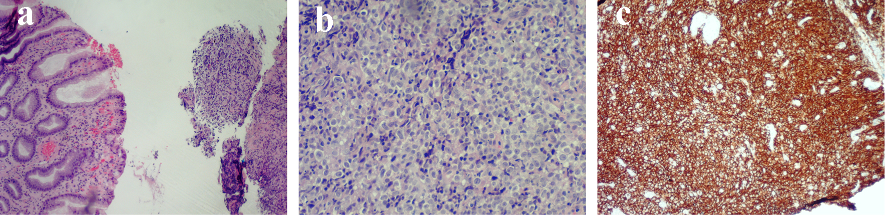
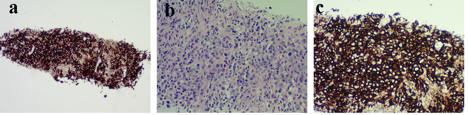
Figures