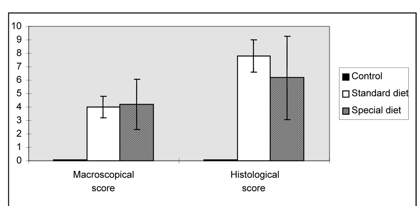
Figure 1. Morphological lesion scores for the intestinal damage induced by indomethacin. The results are shown as the mean ± SD.
| Gastroenterology Research, ISSN 1918-2805 print, 1918-2813 online, Open Access |
| Article copyright, the authors; Journal compilation copyright, Gastroenterol Res and Elmer Press Inc |
| Journal website http://www.gastrores.org |
Original Article
Volume 8, Number 5, October 2015, pages 265-273
Effects of Glutamine and Omega-3 Fatty Acids on Erythrocyte Deformability and Oxidative Damage in Rat Model of Enterocolitis
Figures
Table
| Groups | Baseline weight | Final weight | Difference in body weight |
|---|---|---|---|
| The results are shown as the mean ± SD. | |||
| Control (n = 5) | 235.8 ± 19.3 | 248.9 ±17.9 | 13.1 |
| Standard diet (n = 7) | 244.5 ± 8.7 | 174.3 ± 6.8 | -3.4 |
| Special diet (supplemented with Gln + omega-3 FA) (n = 7) | 270 ± 12.1 | 266.2 ± 10.3 | -4.2 |