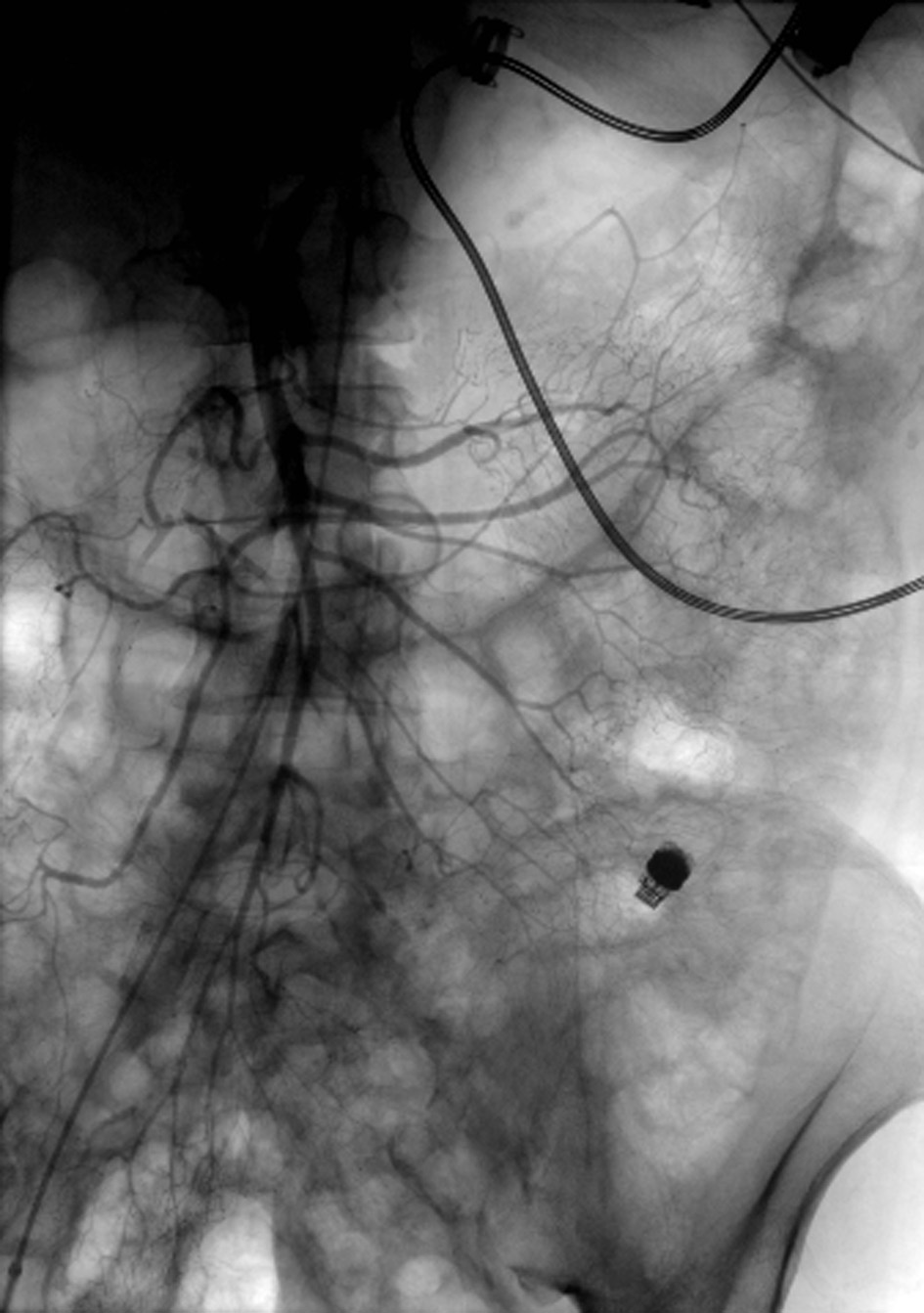
Figure 1. Angiogram of the superior mesenteric artery without extravasation of contrast. Notice the LVAD with driveline in the left upper quadrant and the PillcamSB in the left lower quadrant.
| Gastroenterology Research, ISSN 1918-2805 print, 1918-2813 online, Open Access |
| Article copyright, the authors; Journal compilation copyright, Gastroenterol Res and Elmer Press Inc |
| Journal website http://www.gastrores.org |
Case Report
Volume 8, Number 6, December 2015, pages 309-312
Safety of Deep Enteroscopy and Capsule Endoscopy in LVAD Patients: Case Report and Literature Review
Figures
Table
| Publication author, year | Capsule study | Enteroscopy | Direct complications of capsule study or enteroscopy |
|---|---|---|---|
| DBE: double balloon enteroscopy; min: minutes; push: push enteroscopy; SBE: single balloon enteroscopy; spiral: spiral enteroscopy. | |||
| Girelli et al, 2006 [9] | 1 | None | |
| Seow and Zimmerman, 2006 [10] | 1 | One push | None |
| Fenkel et al, 2007 [11] | 1 | None | |
| Daas et al, 2008 [12] | 1 | One push | None |
| Bechtel et al, 2010 [13] | 1 | None | |
| Decker et al, 2010 [5] | 1 | One DBE | None |
| Stern et al, 2010 [4] | 3 | None | |
| Demirozu et al, 2011 [14] | Two push | None | |
| Elmunzer et al, 2011 [6] | 13 | Four SBE/DBE; three push | None |
| Huang et al, 2012 [15] | 1 | None | |
| Kushnir et al, 2012 [2] | 5 | One push | Duodenal ulcer perforation after three episodes of electrocautery |
| Harris et al, 2013 [8] | 14 | Two patients with brief interruptions (< 2 min) in capsule imaging | |
| Sarosiek et al, 2013 [7] | Thirteen spiral/SBE/DBE | None | |
| Shrode et al, 2014 [3] | 20 | Ten SBE/DBE | None |
| Total | 62 | Eight push; 28 spiral/SBE/DBE | One duodenal ulcer perforation Two disruptions of capsule imaging |