Gastroenterology Research, ISSN 1918-2805 print, 1918-2813 online, Open Access
Article copyright, the authors; Journal compilation copyright, Gastroenterol Res and Elmer Press Inc
Journal website http://www.gastrores.org

Case Report

Volume 8, Number 2, April 2015, pages 197-200


A Rare Case of an Intraductal Papillary Mucinous Neoplasm of Pancreas Fistulizing Into Duodenum With Adult Polycystic Kidney Disease

Figures

Figure 1.
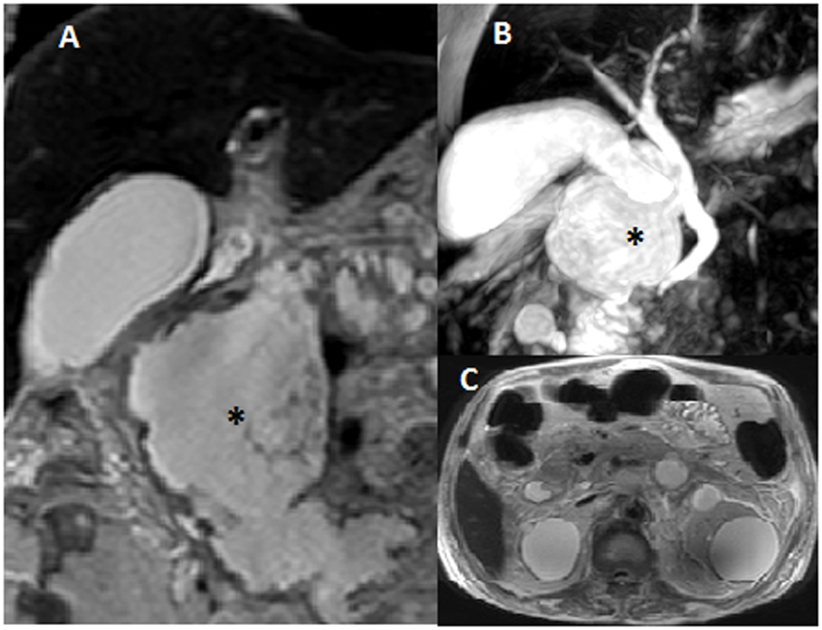
Figure 1. Magnetic resonance cholangiopancreatography with MRI abdomen with pelvis showed large 8 × 5 × 5 cm cystic irregular collection (asterisk) with solid component and septations involving head and body of the pancreas, almost replacing them (A, B). Bilateral multiple cysts in kidneys were also noted (C).
Figure 2.
Figure 2. Endoscopic ultrasound revealed ill defined cystic lesion in the head of the pancreas with some solid component (A, B, C). EUS guided needle aspiration was taken (B). Pancreatic duct was dilated (7 mm) (asterisk) (D). The lesion was communicating with the second part of duodenum.
Figure 3.
Figure 3. Esophagoduodenoscopy showing an fistulous opening in the second part of duodenum just distal to the papilla. Opening was discharging mucus and debris (A). Tumor cavity with solid component was seen from the fistulous opening (B).