Gastroenterology Research, ISSN 1918-2805 print, 1918-2813 online, Open Access
Article copyright, the authors; Journal compilation copyright, Gastroenterol Res and Elmer Press Inc
Journal website http://www.gastrores.org

Case Report

Volume 8, Number 2, April 2015, pages 193-196


Primary Pancreatic Head Tuberculosis: Great Masquerader of Pancreatic Adenocarcinoma

Figures

Figure 1.
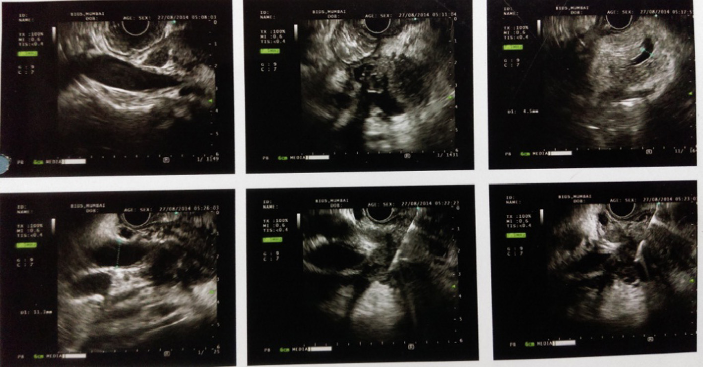
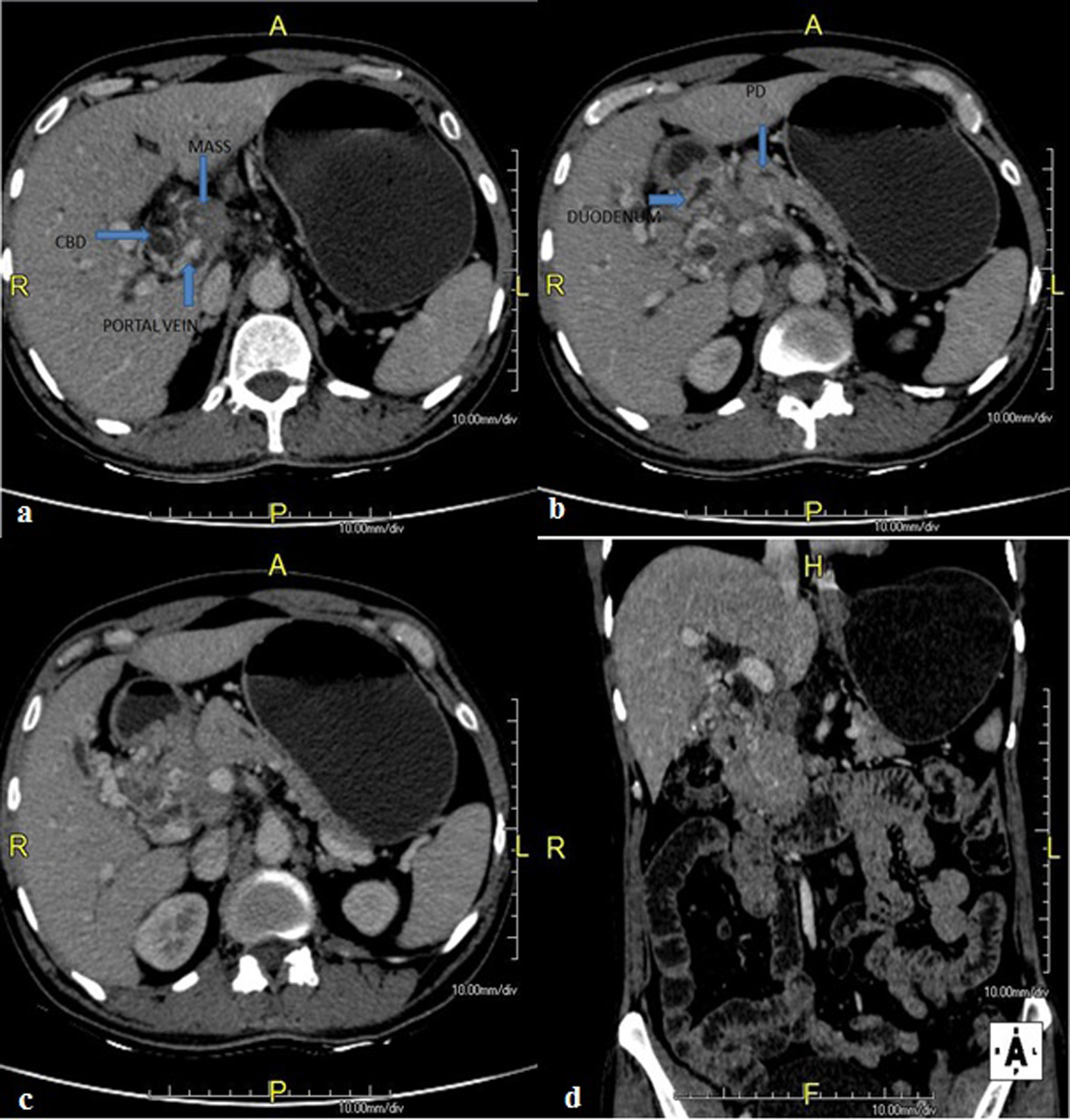
Figure 1. CECT scan abdomen showing hypodense lesion in pancreas head with bulky head with multiple discrete lymph nodes, largest measuring 1.3 × 1.2 cm with compression of main portal vein with presence of multiple collaterals. Pancreatic duct was dilated with 6 mm diameter in head region. Double duct sign was present.
Figure 2.
Figure 2. Endoscopic ultrasound (EUS) showing hypoechoic lesion in pancreas head causing dilation of CBD (11.3 mm) and PD (5.3 mm) with compression of portal vein and multiple peripancreatic and perilesional nodes present.
Figure 3.
Figure 3. Fine needle aspiration cytology showing (A-C) epithelioid cell granulomas with Langhans giant cells with no evidence of malignancy. (D) Acid-fast bacillus stain was positive.